

Executive Overview
For generations, rheumatologists, immunologists, and patients alike have wrestled with one of the most persistent enigmas in modern medicine: why does rheumatoid arthritis (RA) meticulously target specific joints while leaving others completely untouched? For decades, conventional medical wisdom attributed this selective devastation almost entirely to the vagaries of the human immune system, post-natal mechanical stress, or random systemic inflammatory flare-ups.
However, a landmark study published in Nature Immunology by a multidisciplinary team of researchers at the Kennedy Institute of Rheumatology at the University of Oxford, in collaboration with the University of Birmingham, University College London, and the Diamond Light Source, has upended this long-standing paradigm. The researchers have discovered that the seeds of joint vulnerability are sown long before a person takes their first breath.
According to the study, titled "The embryonic origins of site-specific arthritis," certain human joints are biologically preprogrammed for vulnerability to inflammatory conditions during embryonic development. By mapping developing human joints using cutting-edge single-cell sequencing, advanced spatial image analysis, and high-resolution 3D X-ray scanning, the research team revealed that structural and cellular differences between disease-prone joints and immune-spared joints are already firmly established before birth.
This revelation shifts the narrative of autoimmune disease pathogenesis. It suggests that the local microenvironment of a joint—complete with specialized connective tissue cells and distinct structural geometries laid down in the womb—serves as a primary architect in determining where rheumatoid arthritis takes hold decades later. This profound shift in understanding not only redefines our comprehension of autoimmune pathology but also opens up entirely novel therapeutic horizons aimed at intercepting disease before symptoms ever manifest.
Detailed Chronology: How the Breakthrough Unfolded
To untangle a puzzle that has stymied researchers for decades, the Oxford-led team had to pioneer a novel methodological approach. Studying adult joints ravaged by chronic inflammation often obscures the original inciting factors, as secondary tissue damage, chronic scarring, and heavy immune cell infiltration mask the primary cellular landscape. Consequently, the researchers turned their attention to human embryonic and fetal development, creating a chronological blueprint of how joints form.
Step 1: Mapping the Developing Joint Landscape
Using high-resolution single-cell RNA sequencing alongside advanced image analysis, the investigators charted the cellular dynamics of human finger joints at various stages of prenatal development. This allowed them to capture the architecture of the joint at its inception, providing a level of anatomical and cellular clarity previously unattainable in adult tissue studies.
During these early developmental windows, the researchers observed that developing joints are predominantly populated by structural cells rather than immune cells. These foundational populations include early fibroblasts and chondrocytes (cartilage-forming cells). By tracking the signaling pathways that direct these precursor cells to differentiate into specialized lineages, the team gained critical insight into how the synovial lining—the delicate membrane that lubricates joints and nourishes cartilage—is initially formed.
Step 2: Comparing Vulnerable and Resilient Joints
To pinpoint why rheumatoid arthritis selectively targets certain areas, the team chose a comparative model: two distinct types of finger joints that exhibit polarized patterns of disease susceptibility in adult life.
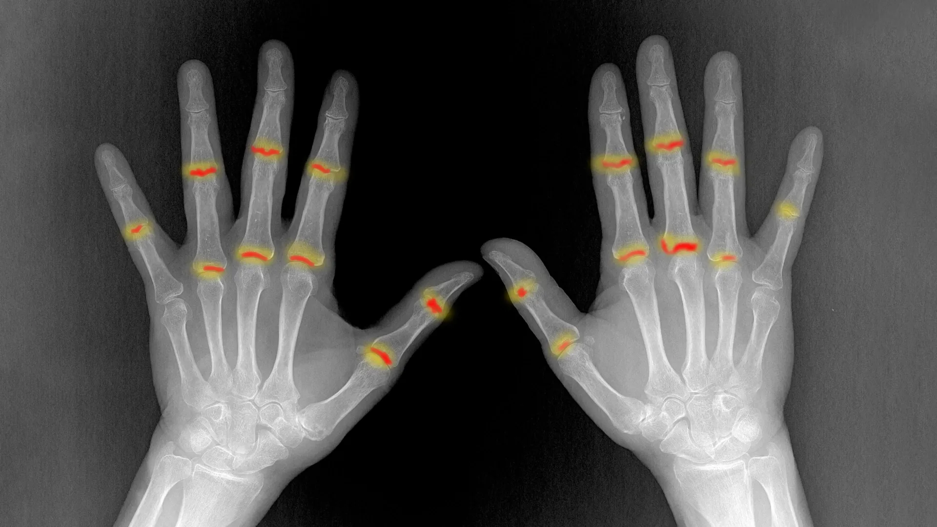
- Proximal Interphalangeal (PIP) joints: Located closer to the hand, these joints are notoriously and frequently targeted by rheumatoid arthritis.
- Distal Interphalangeal (DIP) joints: Located near the fingertips, these joints are generally spared from the ravages of RA.
By holding these two joint types side-by-side through developmental tracking, the researchers isolated the physical and cellular divergences that emerge as the embryo matures.
Step 3: Discovering Prenatal Discrepancies
The comparative analysis yielded striking results. Long before birth, the PIP joints (vulnerable) and DIP joints (resilient) were already morphologically and cellularly distinct. The future RA-prone PIP joints possessed a significantly larger volume of synovial tissue. Furthermore, this synovial tissue featured a dense accumulation of a specialized subpopulation of connective tissue cells known as PI16-positive (PI16+) fibroblasts.
Using high-resolution 3D X-ray imaging facilities at the Diamond Light Source—the UK’s national synchrotron science facility located at the Harwell Science and Innovation Campus—the team confirmed that these structural and volumetric differences persisted into maturity. The spatial arrangement of the synovium in PIP joints was fundamentally different from that of DIP joints, suggesting that prenatal developmental programs dictate the structural topography upon which future pathology is built.
Supporting Context & Metrics: The Cellular Mechanics of Vulnerability
To fully grasp the significance of these findings, it is necessary to examine the pathology of rheumatoid arthritis and the specific cellular actors identified in the Oxford study.
The Anatomy of an Autoimmune Assault
Rheumatoid arthritis is a chronic, systemic autoimmune disease affecting approximately 1% of the global population. Unlike osteoarthritis, which is primarily a degenerative wear-and-tear condition, RA occurs when the body’s immune system erroneously targets the synovium—the specialized membrane lining the joint capsule.
In a healthy individual, the synovium produces synovial fluid, which lubricates the joint and minimizes friction during movement. In a patient with rheumatoid arthritis, immune cells flood the synovium, causing it to become chronically inflamed, swollen, and painful (a condition known as synovitis). Over time, this inflamed tissue transforms into an aggressive, tumor-like mass called a pannus. The pannus invades and erodes adjacent cartilage and bone, leading to permanent joint deformity, loss of function, and severe disability.
The Role of PI16+ Fibroblasts
At the heart of the Kennedy Institute’s discovery are fibroblasts—versatile connective tissue cells that maintain the structural integrity of connective tissues. Within the synovium, specialized fibroblasts play a vital role in maintaining joint homeostasis. However, in the context of arthritis, these cells can undergo pathological reprogramming, driving aggressive inflammation and joint destruction.
The research team identified that PI16+ fibroblasts (fibroblasts expressing the protein CD169/PI16) were heavily enriched in the prenatally vulnerable PIP joints compared to the spared DIP joints. These specialized cells were strategically clustered around blood vessels and at anatomical junctures where tendons and ligaments anchor into adjacent tissues—precisely the high-stress mechanical zones where rheumatoid arthritis lesions frequently initiate.
When the researchers exposed these various fibroblast populations to inflammatory signaling molecules in vitro, they discovered that PI16+ fibroblasts behaved differently than their PI16- counterparts. While both populations mounted a general pro-inflammatory response, the PI16+ cells exhibited unique, highly specialized alterations in biological pathways tied directly to immune regulation and extracellular matrix organization.
Analytical Metrics and Methodological Rigor
- Multi-Platform Approach: The study integrated single-cell transcriptomics, spatial transcriptomics, and high-resolution 3D synchrotron X-ray micro-tomography.
- Institutional Collaboration: Spearheaded by the Kennedy Institute of Rheumatology at the University of Oxford, the project integrated cross-disciplinary expertise from the University of Birmingham, University College London, and the Diamond Light Source.
- Primary Funding: The research was generously funded and supported by the Medical Research Council (MRC), underscoring its high priority within the UK biomedical research landscape.
Official Statements and Expert Perspectives
The implications of the study have resonated deeply across the international rheumatology community. Leading voices behind the research have emphasized both the foundational nature of the discovery and its long-term clinical potential.
Professor Christopher Buckley, Kennedy Professor of Translational Rheumatology at the University of Oxford and senior author of the study, reflected on the historical context of the research:
"For decades we have known that rheumatoid arthritis selectively targets particular joints, but one of the great unanswered questions is why? Our findings suggest that the answer lies not only in the immune system but also in the tissues themselves. The cellular and structural characteristics established during development may help determine where inflammation takes hold later in life."
Dr. Buckley’s remarks underscore a fundamental paradigm shift: rather than viewing autoimmune diseases purely as an immune system malfunction that happens to land on random body parts, medicine must now account for the localized "soil" (the joint tissue environment) as much as the "seed" (the immune system).
Dr. Sarah Davidson, a postdoctoral researcher at the Kennedy Institute and one of the lead authors of the study, elaborated on the micro-anatomical findings:
"We found that joints commonly affected by rheumatoid arthritis already contain distinct cellular populations before birth. PI16+ fibroblasts were enriched in vulnerable joints and responded differently to inflammatory signals. Their location and behavior suggest they could help shape where disease develops."
Dr. Davidson’s insights point toward a future where physicians can identify individuals at risk based on subtle structural and cellular phenotypes long before clinical signs of arthritis manifest.
Future Outlook: Translating Embryology into Clinical Therapeutics
The publication of "The embryonic origins of site-specific arthritis" in Nature Immunology marks not a conclusion, but a vibrant beginning for a new wave of translational research. By establishing that joint vulnerability is hardwired during embryonic development, scientists can now pursue novel avenues for prevention, diagnosis, and treatment.
1. Precision Therapeutics Targeting Synovial Subpopulations
Current treatments for rheumatoid arthritis—ranging from traditional disease-modifying anti-rheumatic drugs (DMARDs) to advanced biologic agents and JAK inhibitors—tend to focus on dampening the systemic immune response. While these therapies have revolutionized patient care, they often carry broad immunosuppressive side effects and do not work equally well for all patients.
By identifying PI16+ fibroblasts as key cellular players in disease-prone joints, researchers can now design precision therapies that specifically target these pathological fibroblast subsets without broadly shutting down the patient’s immune system. Restoring normal, protective behavior to synovial fibroblasts could halt the destructive cascade of arthritis at its very source.
2. Early Risk Stratification and Predictive Diagnostics
Understanding that structural and cellular vulnerabilities are present before birth opens up exciting possibilities for early risk assessment. While screening unborn children is neither necessary nor practical for a disease that typically manifests in middle age, advanced imaging techniques and biomarker profiles could eventually be used to assess joint vulnerability in patients with a strong genetic predisposition or early, undifferentiated joint pain.
By identifying which specific joints are most at risk in a given patient, clinicians could deploy targeted, localized interventions (such as site-specific prophylactic treatments) to prevent inflammation from ever taking hold.
3. Regenerative Medicine and Joint Engineering
As scientists continue to map the developmental signals that dictate how synovial linings and joint structures form, these insights will inevitably feed into the fields of regenerative medicine and tissue engineering. Understanding how nature builds a resilient joint versus a vulnerable one could inform the design of bioengineered joint replacements, cartilage repairs, and regenerative therapies for osteoarthritis and trauma-induced joint injury.
Conclusion
The work conducted at the Kennedy Institute of Rheumatology bridges developmental biology and immunology in an unprecedented way. By looking backward to the earliest moments of human embryogenesis, researchers have illuminated a clear path forward for conquering one of rheumatology’s most stubborn mysteries. As these findings move from the bench to the clinical trial pipeline, millions of patients suffering from inflammatory joint diseases may soon benefit from therapies informed by the very blueprint of human creation.