

Executive Overview
Long before a human infant draws its first breath, an extraordinary, hyper-orchestrated symphony of cellular decisions constructs the most complex object known in the universe: the human brain. At the heart of this pre-natal architectural marvel are radial glia—specialized, highly dynamic stem cells that serve as the master builders of the cerebral cortex. This sheet of neural tissue is responsible for humanity’s highest-order capabilities, including abstract thought, autobiographical memory, language processing, and complex reasoning.
While scientists have long recognized the pivotal role of radial glia, the exact mechanics governing how these master stem cells decide when to divide, what specific types of neurons to spawn, and how to fuel their rapid transformations have remained an elusive frontier. Now, two monumental, concurrent studies published in the premier journals Cell and Science by a collaborative team of researchers at the University of California, Los Angeles (UCLA) have pierced this veil of mystery.
Led by Dr. Aparna Bhaduri, an assistant professor of biological chemistry at the David Geffen School of Medicine at UCLA, these investigations demonstrate that radial glia do not operate in a vacuum. Instead, their developmental decisions are dictated by two surprising external inputs: internal metabolic processing pathways and direct, physical cellular signaling from deep within the developing brain.
By marrying cutting-edge human tissue analysis with advanced laboratory-grown brain organoids and "assembloids," the UCLA research teams have charted an unprecedented metabolic atlas and uncovered a previously unknown physical dialogue between the thalamus and cortical stem cells. Furthermore, these revelations have cast new light on how disruptions in these pathways may lay the structural groundwork for neurodevelopmental conditions, including autism spectrum disorder, as well as aggressive brain cancers where radial glia-like cells inappropriately re-emerge.
Detailed Chronology of Discovery: Unlocking the Secrets of Radial Glia
To understand the magnitude of the recent breakthroughs, one must first appreciate the evolutionary prominence of radial glia. These cells are widely believed to drive the unusually massive expansion of the human cerebral cortex compared to our evolutionary relatives and other mammalian species. Though the vast majority of these stem cells disappear or differentiate before birth, their genetic and behavioral signatures can later resurface in malignant brain tumors—a phenomenon that continues to baffle and challenge oncologists and neuroscientists alike.
"Radial glia are the coolest cells that have ever existed," asserts Dr. Bhaduri, who holds appointments at the UCLA Broad Stem Cell Research Center and the UCLA Health Jonsson Comprehensive Cancer Center. "They’re really key to making us human. But they’re also at the center of many neurodevelopmental and neuropsychiatric disorders, as well as cancer—so understanding how they make their decisions is one way to start understanding how those conditions arise."
The path to decoding these cellular choices unfolded across two distinct yet complementary research tracks, utilizing cutting-edge laboratory models that have revolutionized developmental biology over the past decade.
Phase I: Metabolism as an Active Master Regulator (Cell)
Historically, cellular metabolism—the chemical processes by which cells convert nutrients into energy and building blocks—was viewed by developmental biologists as a passive background support system. It was thought to supply the raw fuel required for growth, but little more. The first of the new UCLA studies, published in Cell, shatters this long-standing assumption.
Co-led by Dr. Bhaduri’s laboratory and the lab of Dr. Heather Christofk, with co-first authors Jessenya Mil and Jose Soto, the research team set out to construct a high-resolution, detailed metabolic map of the developing human cortex. To achieve this, the investigators analyzed donated human embryonic and fetal tissue alongside sophisticated brain organoids—three-dimensional cellular structures grown from human stem cells in vitro that mimic key aspects of early brain architecture.
The resulting metabolic atlas pointed to an unexpected conclusion: metabolism actively dictates cell fate. Specifically, the researchers discovered that radial glia rely heavily on a specific biochemical pathway known as the pentose phosphate pathway. This metabolic route utilizes glucose not merely for cellular respiration and basic survival, but to synthesize the essential molecular building blocks required by rapidly dividing, highly proliferative cells.
When the research team experimentally manipulated this environment—either by lowering the availability of glucose or by chemically interfering with the pentose phosphate pathway—the stem cells dramatically altered their production output. Rather than maintaining their standard developmental trajectory, they began generating an overabundance of inhibitory neurons and alternative cell types that typically do not emerge until much later in gestation.
"What was surprising is that metabolism isn’t just a passive thing that happens in the background," Dr. Bhaduri explains. "It can really control how stem cells make decisions."
This discovery bridges a critical knowledge gap in developmental biology, offering a concrete biochemical link between environmental factors—such as maternal nutrition, gestational metabolic disorders, and systemic inflammation—and their potential long-term impacts on fetal neurodevelopment.
Phase II: The Thalamic Touch (Science)
While the Cell study examined internal metabolic programming, the second investigation, published in Science and led by first author Claudia Nguyen, shifted focus to external, long-range communication networks within the developing brain.
For years, neuroscientists have documented that neurons residing in the thalamus—a deep-seated sensory relay structure located near the center of the brain—extend exceptionally long, wire-like projections toward the developing cortex. These neural fibers eventually form intricate synaptic connections with specific cortical neurons, processing sensory information ranging from vision to touch. However, detailed anatomical studies of human fetal tissue revealed a curious biological anomaly: these thalamic projections arrive at the cortex long before those functional synaptic networks are scheduled to be established.
This chronological discrepancy raised a fundamental question: Why do these neural fibers complete their long journey so early?
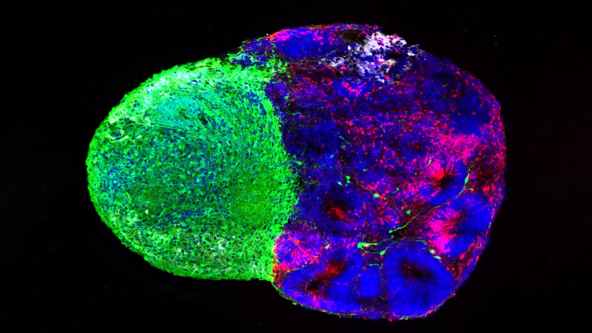
Utilizing human stem cell-derived brain "assembloids"—complex models formed by fusing two or more distinct types of organoids to study inter-regional communication—the UCLA team discovered the answer. Contrary to the prevailing assumption that neural fibers wait silently for their targets to mature, the thalamic projections physically reach out and directly touch radial glia while the cerebral cortex is still in its infancy.
This physical contact acts as an immediate developmental trigger. When the thalamic fibers touch the radial stem cells, they alter the cells’ behavior, prompting them to generate a higher proportion of excitatory neurons. These are the primary signal-carrying neurons of the cortex, and their production is especially pronounced in the upper layers of the human brain—the regions that expanded most dramatically during human evolution.
"We already knew that these projections influence how the cortex develops," notes Dr. Bhaduri. "What we specifically found is that this influence comes through an actual physical connection between the projections and the radial glia—a point of contact that just hasn’t been identified before, and one that very likely does not exist in rodents."
Supporting Context, Metrics, & Molecular Intersections
The convergence of metabolic signaling and physical neural contact illustrates a profound biological truth: human brain development is a masterclass in multi-layered communication. Radial glia do not execute an internal genetic script in isolation; they are continuously buffeted, shaped, and instructed by their immediate microenvironment.
The Genetic Link to Autism: NRXN1
To demonstrate the clinical and translational relevance of these discoveries, the UCLA team investigated what happens when this delicate cellular dialogue is disrupted. They focused their attention on NRXN1, a gene that encodes neurexin-1, a cell-adhesion molecule well-known for helping neurons anchor and form stable synaptic connections with one another. Mutations in the NRXN1 gene have long been strongly correlated with an increased risk for autism spectrum disorder (ASD) and other neurodevelopmental conditions.
By engineering brain assembloids from patient-derived stem cells carrying a targeted NRXN1 mutation, the researchers observed a stark departure from normal development. In these mutated models, the signals transmitted by the thalamic projections behaved aberrantly compared to those generated by healthy, unaffected cells.
This cellular miscommunication disrupted the finely tuned balance between the pool of active stem cells and the rate at which they generated downstream neurons. By revealing how a gene linked to autism alters early physical interactions between brain regions, this research provides investigators with a human-relevant model to study how microscopic disturbances during early embryogenesis can cascade into complex neurobehavioral conditions later in life.
Official Statements and Perspective
The implications of these dual studies extend far beyond basic developmental biology, offering a unified framework for viewing brain formation, pathology, and evolution.
Dr. Bhaduri emphasizes that these findings challenge researchers to re-evaluate traditional paradigms in neurobiology:
"Ultimately, these studies give us a glimpse under the hood of how these cells make decisions. Understanding those decisions is a first step toward understanding normal brain development, disease vulnerability and, potentially, how similar stem-cell programs operate in brain cancer."
The research also underscores the technological leap represented by modern organoid and assembloid systems. Only a decade ago, studying uniquely human neural stem cell dynamics in a laboratory setting was virtually impossible, as rodent models fail to replicate the vast evolutionary expansions and structural nuances of the human cerebral cortex. Today, these advanced in vitro platforms allow scientists to ask complex, mechanistically precise questions about human-specific biology that were once entirely out of reach.
Future Outlook: Toward Precision Neurobiology and Oncology
As the scientific community digests the insights provided by the UCLA Broad Stem Cell Research Center and the UCLA Health Jonsson Comprehensive Cancer Center, the path forward points toward several high-impact avenues of investigation:
- Translational Metabolism: Future studies will examine whether targeted dietary interventions or metabolic modulators can mitigate the developmental impacts of gestational metabolic syndromes and maternal nutritional deficits.
- Decoding Neurodevelopmental Disorders: By refining patient-derived assembloid models featuring mutations in genes like NRXN1, researchers hope to pinpoint specific temporal windows during pregnancy where therapeutic interventions might rescue or correct aberrant neural circuitry.
- Cancer Stem Cell Therapeutics: Because malignant brain tumors co-opt the survival and division programs of embryonic radial glia, understanding the precise metabolic and physical triggers that govern these stem cells could reveal novel vulnerabilities for targeted cancer therapies.
Through the integration of high-resolution metabolic mapping and advanced human tissue modeling, science is steadily decoding the intricate cellular choreography that builds the human mind—opening unprecedented doors to treating the disorders that afflict it.
This groundbreaking research was generously supported by a robust coalition of funding bodies and philanthropic organizations, including the National Institutes of Health (NIH), the National Science Foundation, the Brain & Behavior Research Foundation, the Alfred P. Sloan Foundation, the Rose Hills Foundation, the Esther A. & Joseph Klingenstein Fund, the Simons Foundation, the Chan Zuckerberg Initiative, the NIH BRAIN Initiative Cell Atlas Network, the International Foundation for Ethical Research, the UCLA Broad Stem Cell Research Center’s Stem Cell Research Training Program, the UCLA Health Jonsson Comprehensive Cancer Center, and the UCLA Broad Stem Cell Research Center Ablon Scholars Program.