

Executive Overview
Nearly a century after a breakthrough in light microscopy unlocked the internal architectures of living cells, a team of physicists at the University of California, Berkeley, has achieved a monumental parallel in the realm of electron microscopy. By fusing quantum optics with sub-atomic imaging, researchers have developed a revolutionary device known as a laser phase plate. This breakthrough directly addresses one of structural biology’s most persistent bottlenecks: the inability of conventional cryo-electron microscopy (cryo-EM) and cryo-electron tomography (cryo-ET) to clearly resolve small proteins and complex, crowded cellular interiors.
Traditional cryo-EM has transformed structural biology over the past decade, earning a Nobel Prize in 2017 and streamlining the drug discovery pipeline by eliminating the arduous requirement to crystallize biomolecules. Yet, a glaring limitation remained: most human and animal proteins are simply too small or generate too little signal-to-noise contrast to be imaged effectively. Because macromolecules like water-based biological components are largely transparent to electron beams, identifying them within the dense cellular milieu has historically resembled searching for a single leaf in a vast, tangled forest.
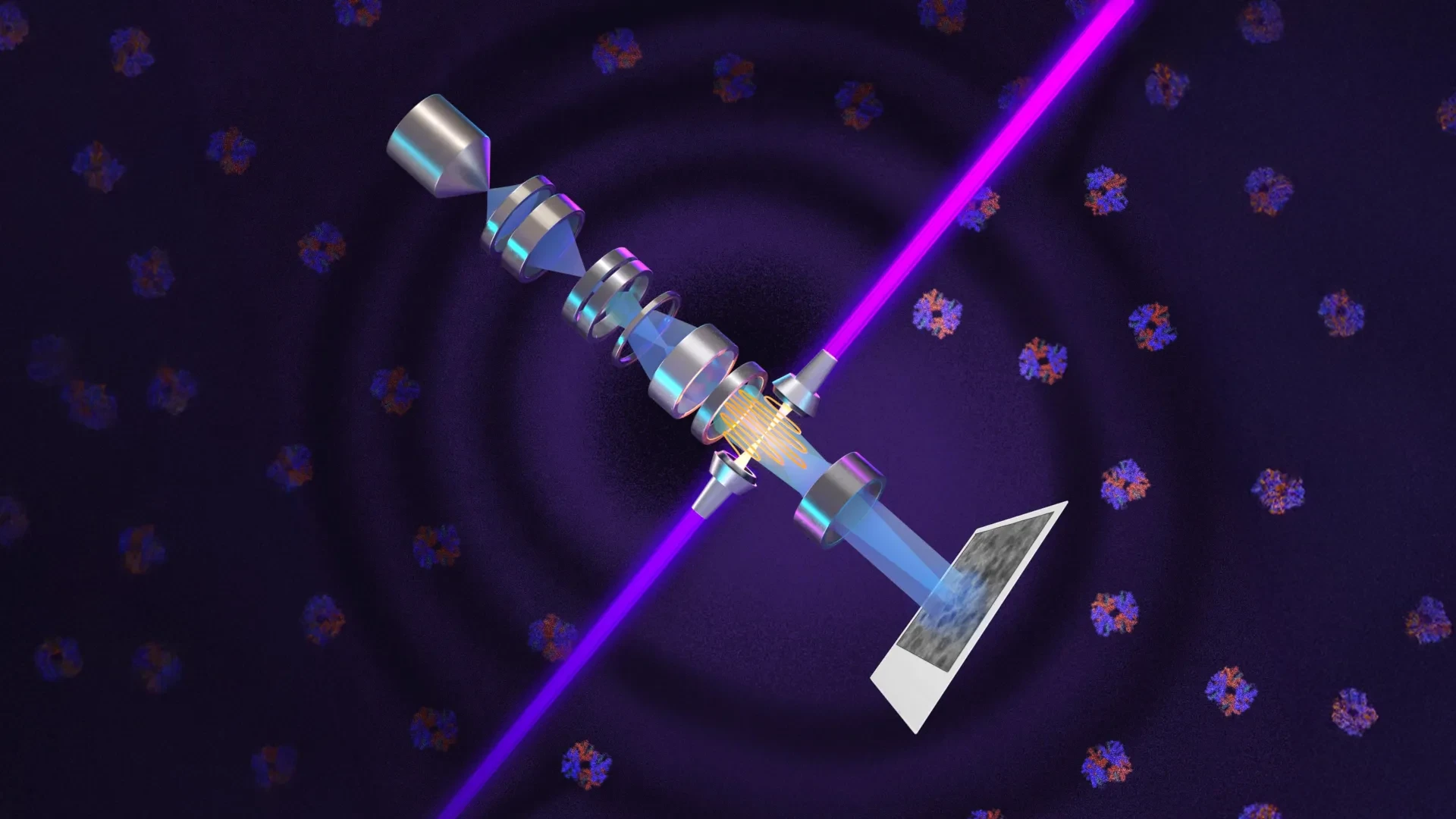
By deploying what is currently the world’s most intense focused continuous-wave laser, UC Berkeley physicists—in collaboration with researchers at the Chan Zuckerberg Biohub and instrument manufacturer Thermo Fisher Scientific—have bypassed this physical barrier. Named "Theia" after the ancient Greek Titaness of light and radiance, this custom-built, Formula 1-grade microscope alters the phase of an electron beam rather than merely illuminating a specimen. The result is a dramatic leap in image contrast and resolution. This technological triumph not only pushes the boundaries of molecular visibility down to previously unachievable size thresholds—dropping from a previous lower limit of roughly 70 kilodaltons down to 50 kilodaltons, with sights set on 17 kilodaltons—it promises to turn the dynamic, in-situ observation of living cellular machinery from a distant theoretical goal into an empirical reality.
Detailed Chronology: A Century of Phase Innovation
The conceptual lineage of the laser phase plate stretches back nearly a hundred years, tracing a path through fundamental physics, engineering roadblocks, and decades of relentless pursuit.
1930: The Birth of Phase Contrast
In 1930, Dutch physicist Frits Zernike observed that light passing through transparent biological specimens undergoes more than a simple attenuation of brightness; it also experiences minute phase shifts—slight delays in the timing of its wave peaks caused by internal scattering. Because the human eye cannot directly register these phase modifications, Zernike devised an optical trick: shifting the phase of unscattered background light by 90 degrees. This caused subtle structural features to stand out with crisp brightness against their backgrounds. This breakthrough birthed phase-contrast microscopy, earning Zernike the 1953 Nobel Prize in Physics and permanently expanding humanity’s window into cellular biology.
Early 1940s – 2010: The Electron Beam Dilemma
Almost as soon as phase contrast was validated for light, physicists wondered if the principle could be translated to electron microscopes, which use beams of electrons to achieve magnifications 10,000 times greater than conventional optics. However, early efforts stalled. Devices engineered to alter electron phase paths invariably degraded the beam, compromised resolution, or introduced catastrophic instabilities.
The theoretical turning point arrived in 2010, when UC Berkeley physicist Holger Müller and Robert Glaeser, now professor emeritus of molecular and cell biology, published a seminal proposal: utilizing an intense laser field to impart the necessary 90-degree phase shift onto an electron beam without sacrificing its intensity or coherence.
2010 – 2025: The 15-Year Engineering Odyssey
What looked elegant on paper required fifteen years of painstaking engineering. The core hurdle was generating an optical field of unprecedented intensity—concentrating enough light into a microscopic focal point to interact meaningfully with high-energy electrons.
Müller’s team eventually solved this by trapping a continuous laser beam inside a specialized spherical optical cavity lined with high-reflectivity mirrors. As the light bounced more than 10,000 times within the micro-chamber, its energy compounded exponentially into an intensely focused spot. With financial backing from the National Institutes of Health and later the Chan Zuckerberg Biohub, the team integrated this system into a prototype electron microscope, validating the concept before scaling it up into a customized Thermo Fisher Krios instrument.
2026: Validation and Public Debut
In early 2026, Müller’s team published their definitive technical findings and structural reconstructions in the journal Science, while Biohub researchers simultaneously released preprints detailing a sophisticated dual-laser configuration designed to eliminate optical distortions and prevent component degradation. These milestones formally ushered in the era of laser-enhanced electron microscopy.
Supporting Context & Metrics: Overcoming Physical Limits
To appreciate the gravity of the UC Berkeley breakthrough, one must examine the fundamental physical constraints governing macromolecular imaging.
+-------------------------------------------------------------------------+
|                  THE EVOLUTION OF BIOLOGICAL IMAGING                    |
+-------------------------------------------------------------------------+
| 1930s: Light Phase Contrast (Frits Zernike)                             |
|   └─ Solved cell transparency via 90-degree light phase shifting.       |
|                                                                         |
| 1970s-2010s: Cryo-EM & Cryo-ET (Glaeser et al., 2017 Nobel Prize)       |
|   └─ Froze samples to prevent radiation damage; bypassed crystallization|
|      but struggled with small proteins (<70 kDa) and dense cellular noise.|
|                                                                         |
| 2026: Laser Phase Plate / Theia Microscope (UC Berkeley & Biohub)       |
|   └─ Uses a 75-kilowatt continuous-wave laser focus to shift electron   |
|      phases directly, pushing imaging limits down to 50 kDa and beyond. |
+-------------------------------------------------------------------------+
The Dalton Barrier and the Human Proteome
Protein mass is measured in daltons (Da), where one dalton equals one-twelfth the mass of a carbon-12 atom. Prior to the advent of the laser phase plate, conventional cryo-EM faced a strict lower threshold of approximately 70 kilodaltons (kDa). This barrier represented a major scientific crisis because proteins below 70 kDa account for roughly 90% of the human proteome. Critical regulatory enzymes, signaling molecules, and structural units remained stubbornly invisible or obscured by background noise.
Powering the Invisible
The laser system developed at Berkeley achieves densities that defy everyday comprehension:
- Circulating Power: 75 kilowatts of continuous-wave laser light.
- Focal Area: Confined to a cross-section of only a few microns.
- Intensity Comparison: The resulting optical field is more powerful than industrial welding lasers and exceeds the output specifications of military-grade directed-energy systems, creating the brightest continuous laser focus ever recorded.
By running this beam perpendicular to or interacting directly with the electron trajectory, the system imparts a precise phase shift. In performance tests using aldolase and hemoglobin—the latter sitting near the historical floor of conventional cryo-EM capabilities—the laser phase plate drastically enhanced signal-to-noise ratios, successfully resolving structures that were previously washed out in optical noise.
Official Statements and Expert Perspectives
The academic and industrial communities have responded to the breakthrough with high anticipation, emphasizing its disruptive potential for structural biology and drug discovery.
-
Holger Müller, UC Berkeley Professor of Physics & Lead Researcher:
"Cryo-EM has become the new, fastest-growing method for resolving the structure of biological macromolecules, and cryo-ET is expected to show how these molecules work together in their natural, cellular context. But because of signal-to-noise limitations, the majority of human and animal proteins are too small to be analyzed by these methods. The increase in signal-to-noise ratio provided by this laser phase plate is expected to overcome these important limitations."
-
Bridget Carragher, Founding Technical Director of Imaging at Biohub:
"With cryo-ET, we’re looking at small, very complicated cellular material that’s incredibly crowded inside the cell. It’s like a forest of trees, and you’re trying to find one leaf on one tree in there. Cryo-ET needs a dramatic step forward in contrast, so we can start to see what’s going on inside the cell. That’s what the laser phase plate promises to give us."
-
Stephani Otte, Biohub’s Vice President of Imaging Science:
"This technology is a step function change for biology. We are going to be able to see how molecular machines operate inside the living cell, in context, for the first time. What was once invisible will become visible — and that changes everything about how we understand disease."
Future Outlook: The Road to Molecular Transparency
With the viability of the laser phase plate demonstrated on benchmark proteins like hemoglobin and aldolase, the research collaborative has set its sights on even loftier targets.
The immediate technical horizon involves pushing the lower size limit down from 50 kilodaltons to 17 kilodaltons—the molecular weight of myoglobin. Achieving this will require transitioning from the defocused electron beams currently utilized to highly focused electron beams, an adjustment that Müller estimates could double the signal-to-noise contrast once again.
Furthermore, the expansion of cryo-electron tomography (cryo-ET) stands as a transformative frontier. While single-particle cryo-EM studies isolated proteins in frozen solutions, cryo-ET composites images captured from diverse tilt angles to yield three-dimensional, high-resolution maps of entire cellular landscapes. By coupling the unprecedented contrast of the laser phase plate with cryo-ET protocols, scientists will soon be able to watch macromolecular assemblies execute their biological functions in real time, nestled securely within their native cellular neighborhoods.
As institutions like UC Berkeley and Biohub refine these instruments—and as industry leaders like Thermo Fisher Scientific explore pathways toward commercial adaptation—structural biology stands on the precipice of a new golden age. By turning intense light into a master key for the sub-atomic world, researchers are dismantling the final barriers of cellular opacity, illuminating the hidden mechanisms of life itself, and rewriting the future of medical science.