

Executive Overview
In a monumental leap forward for structural biology and biophysics, researchers at the University of Oxford have successfully observed virus-like particles assembling themselves in real time, one molecule at a time. Published in the prestigious journal Nature, this breakthrough shatters decades of scientific limitation, offering an unprecedented, high-resolution view of a fundamental biological process that has historically remained shrouded in mystery.
For generations, scientists understood the end results of viral self-assembly—intricate, highly organized protective protein shells known as capsids—and they could analyze the raw protein building blocks floating in isolation. Yet, the critical transition phase connecting the starting materials to the finished architecture was essentially a black box. Intermediate structures form transiently and in vanishingly small quantities, evading traditional analytical frameworks.
By leveraging pioneering single-molecule measurement technologies, the Oxford research team bypassed these historical roadblocks. They discovered that the astonishing reliability with which complex viral structures form is governed by surprisingly simple physical rules. Rather than stumbling blindly through an astronomical number of potential geometric configurations, proteins navigate a disciplined sequence of molecular checkpoints. Through a sophisticated system of trial and error—facilitated by weak initial bonds that easily break and reform—the building blocks prune away failed pathways until they lock into stable, functional geometries.
This revelation does far more than satisfy fundamental academic curiosity. By mapping the exact kinetic pathways of viral assembly, the discovery paves the way for a paradigm shift in pharmacology. Researchers can now conceptualize novel classes of antiviral drugs designed to deliberately sabotage the assembly process, throwing a wrench into the virus’s construction machinery before it can protect its genetic cargo. Furthermore, these insights will supercharge the engineering of bespoke virus-like particles (VLPs) tailored for cutting-edge vaccines and targeted gene-therapy delivery systems.
Detailed Chronology: Observing the Invisible
To appreciate the gravity of the Oxford breakthrough, one must understand the formidable hurdles associated with capturing viral assembly. Viruses are master structural engineers on the nanoscale. They store, transport, and protect their fragile genetic material—DNA or RNA—inside capsids composed of anywhere from tens to thousands of individual protein subunits.
In many biological contexts, these proteins possess an innate, self-assembling intelligence. Without the aid of ATP, ribosomes, or external mechanical chaperones, the components spontaneously organize into symmetrical, closed geodesic structures. However, this apparent simplicity is deceptive. As individual proteins collide in solution, they face an overwhelming combinatorial explosion of potential bonding arrangements—thousands of ways to connect, the vast majority of which lead to dead ends, malformed aggregates, or thermodynamic traps.
Historically, virologists and biophysicists relied on ensemble-averaging techniques, X-ray crystallography, and cryogenic electron microscopy (cryo-EM). While these methods yield breathtaking structural snapshots of static states, they cannot faithfully chronicle a dynamic, moving process. Transient intermediate states—the fleeting configurations that exist for milliseconds before either progressing or dissolving—are virtually invisible to bulk measurements because they never accumulate in sufficient concentrations.
The Mass Photometry Breakthrough
To conquer this observational barrier, the Oxford team turned to an innovative technology invented within their own institution: mass photometry. This sophisticated analytical technique measures the mass of individual, unlabelled molecules in solution by quantifying the tiny amounts of light they scatter when binding to a surface.
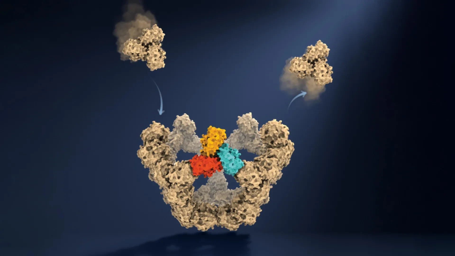
Collaborating across the Department of Chemistry and the Kavli Institute for Nanoscience Discovery, the researchers focused their investigation on an engineered virus-like particle system comprising precisely 60 protein units. By fusing mass photometry with a novel spatial-confinement approach—which traps individual growing particles within an observation zone and tracks them continuously—the team achieved what was previously thought impossible. They filmed the construction process as it unfolded, recording the step-by-step mass accretion of a single viral particle over time.
"This method lets us study the assembly process directly," noted co-first author Dan Loewenthal, a PhD student on the project. "We just take a video!"
What the video revealed challenged prevailing assumptions about molecular kinetics. Rather than executing a chaotic scramble, the proteins displayed an elegant method of trial and error. Initial encounters between protein units generate weak, labile connections. If the geometry is incorrect, these bonds rupture effortlessly, allowing the components to drift apart, reset, and try again.
This inherent flexibility acts as an evolutionary filter. The system continuously discards dead-end configurations while reinforcing successful configurations. The turning point arrives with the formation of the first major structural milestone: a closed pentagonal ring composed of five larger protein building blocks.
Once this pentagonal nucleus forms—marking the end of the notoriously difficult nucleation phase—the thermodynamic landscape shifts dramatically. The assembly gains structural rigidity, acting as a molecular checkpoint. Each subsequent step requires fewer building blocks to achieve stabilization, causing the rate of particle growth to accelerate rapidly until the full 60-protein capsid is triumphantly sealed.
Supporting Context & Metrics
The quantitative rigor underpinning the Oxford study provides a foundational framework for understanding self-assembly mechanics across diverse biological systems. The metrics derived from real-time mass photometry shed light on the thermodynamic efficiencies that make viral replication so lethally effective.
- The 60-Protein Architecture: The study focused on a symmetrical model system consisting of 60 identical protein subunits, mirroring the T=1 icosahedral symmetry found in many small, spherical viruses.
- Combinatorial Complexity: Without guidance, proteins face thousands of theoretical topological configurations upon collision. The discovery reveals that transient intermediate checkpoints reduce this chaotic search space to a highly linear, manageable subset of successful pathways.
- The Nucleation Bottleneck: Quantitative tracking confirmed that nucleation—the initial formation of the first stable multimeric state—is the kinetic rate-limiting step of assembly. Once the initial pentagonal core is locked in, activation energy barriers drop, exponentially increasing the velocity of subsequent subunit recruitment.
- Rebound Dynamics: The measured bond dissociation rates demonstrate that early-stage reversibility is not a flaw in the system, but an essential feature. It prevents kinetic trapping, ensuring that misfolded or misaligned aggregates do not permanently stall the reaction.
These quantitative insights shift our comprehension of self-assembly from qualitative hypothesis to exact mathematical modelling. By directly measuring the binding kinetics and stabilities of intermediate species, researchers can now simulate viral assembly pathways on computers with unprecedented fidelity.
Official Statements from the Research Team
The implications of this breakthrough were articulated by the leading minds behind the Oxford study, highlighting the convergence of advanced physics, chemistry, and molecular biology.
Dr. Roi Asor, co-lead author from the Department of Chemistry and the Kavli Institute for Nanoscience Discovery, emphasized the sheer scale of the biological problem being solved:
"A virus has to solve an extraordinary construction problem. Its components somehow have to find the right arrangement among a huge number of possibilities, without a blueprint or machinery directing the process. We can now watch what happens molecule by molecule and see the physical rules that make it possible."
Elaborating on the mechanism of trial and error, Dr. Asor added:
"The weak interactions give the system room to make mistakes. Most encounters don’t have to be successful: the components can separate and try again. But once enough of them come together in the right closed arrangement, the structure becomes stable, and assembly can move forward. That combination of trial and error followed by locking in successful structures is what makes the process so reliable."
Professor Philipp Kukura, co-lead author and pioneer of mass photometry technology within the Department of Chemistry, reflected on the methodological leap achieved by the team:
"Until now, much of our understanding of how these structures assemble has had to be reconstructed from snapshots or theoretical models. Being able to both quantify the underlying interactions and follow one particle as it grows changes that. We can see the important intermediate structures appear and deduce a detailed model of the construction process."
Looking toward translational applications in medicine, co-author Dr. Jack Tan of the MRC Weatherall Institute of Molecular Medicine and the CAMS-Oxford Institute highlighted the immediate clinical horizons:
"The ability to understand these processes at the molecular level could have important applications in vaccine development and antivirals that disrupt viral assembly."
Future Outlook: Antiviral Pharmacology and Beyond
The publication of these findings in Nature opens a sweeping new frontier in biomedical research, with ripple effects extending far beyond virology.
Next-Generation Antiviral Therapeutics
Traditional antiviral drugs typically target viral enzymes, such as proteases or polymerases, or focus on blocking viral entry into host cells. However, viruses are notoriously adaptable, frequently mutating their enzymatic targets to develop drug resistance.
Capsid assembly inhibitors represent an enticing alternative strategy. Because the physical rules governing protein-protein interactions and geometric closure are deeply conserved constraints rooted in fundamental thermodynamics, viruses cannot easily mutate their way around them without losing structural integrity. By weaponizing the insights gained from Oxford’s mass photometry data, drug developers can now design small molecules or peptides that bind specifically to the intermediate checkpoints or prematurely trigger capsid closure. This effectively forces the virus to assemble incorrectly, encapsidating empty space or destroying its own genome before infection can take hold.
Revolutionizing Vaccine Design and Gene Therapy
Beyond combating pathogenic viruses, the ability to observe and direct self-assembly holds immense promise for biotechnology. Modern vaccine platforms frequently utilize engineered virus-like particles (VLPs) to present foreign antigens to the human immune system in a highly immunogenic, non-infectious format. Similarly, gene therapy vectors—such as engineered adeno-associated viruses (AAVs)—rely on precisely assembled protein shells to deliver therapeutic genes safely into patient tissues.
Currently, the manufacturing yield of functional VLPs and gene vectors can be hampered by inefficient assembly, aggregation, and structural heterogeneity. By understanding the precise physical rules and intermediate checkpoints that govern successful closure, bioengineers can optimize solution environments, mutate specific protein residues, and dramatically improve the yield and purity of therapeutic nanoparticles.
Universal Principles of Biological Self-Assembly
Ultimately, the significance of the Oxford team’s achievement transcends virology. Self-assembly is a universal organizing principle of life itself. From the cytoskeletal filaments that maintain cell shape and direct mitosis, to clathrin-coated vesicles mediating intracellular transport, and macromolecular complexes like ribosomes and proteasomes, nature builds complex biological machinery from simple molecular building blocks.
By proving that it is possible to watch these intricate construction processes play out in real-time, molecule by molecule, the University of Oxford researchers have provided science with a master key. As this methodology is adapted to study other complex biomolecular systems, humanity moves one step closer to reading, mastering, and ultimately mimicking nature’s most sophisticated manufacturing processes.