

Executive Overview
For decades, the medical and scientific communities have grappled with a persistent biological paradox: why do men and women experience obesity, metabolic syndrome, and systemic inflammation so differently? While epidemiological data have long highlighted disparities in fat distribution, cardiovascular risk, and metabolic disease progression between the sexes, the underlying cellular mechanisms have remained frustratingly elusive.
Now, a groundbreaking study led by biomedical scientists at the University of California, Riverside (UCR), has uncovered a fundamental piece of this complex biological puzzle. Published in the journal PNAS Nexus, the research reveals that the immune systems of males and females respond to obesity through starkly divergent pathways governed by unexpected diversity within a specific class of white blood cells: eosinophils.
Traditionally pigeonholed as the body’s frontline defenders against parasitic infections and the primary drivers of allergic reactions and asthma, eosinophils are shedding their monolithic reputation. Utilizing high-resolution single-cell sequencing—a cutting-edge genomic technique capable of parsing gene activity in individual cells—the UCR research team identified six distinct populations of eosinophils residing within adipose (body fat) tissue. More importantly, these populations do not behave identically across sexes.
In female subjects, fat tissue harbors a specialized, protective cohort of eosinophils that engage in sophisticated crosstalk with neighboring macrophages—another critical class of immune cells. This cooperative cellular dialogue fosters an anti-inflammatory, metabolically stable environment. In stark contrast, male subjects consuming a high-fat diet tend to accumulate pro-inflammatory eosinophil variants with diminished protective capabilities. Furthermore, the researchers identified a vital signaling protein, RELM alpha, which acts as a molecular bridge to stabilize the beneficial communication between eosinophils and macrophages.
These findings challenge the long-held biomedical assumption that immune cells within fat tissue are uniform across biological sexes. By demonstrating that the impact of obesity is dictated not merely by the quantity of immune cells, but by their functional heterogeneity and intercellular communication networks, this study paves the way for a paradigm shift in metabolic medicine. As researchers pivot toward human trials and the development of three-dimensional adipose organoids, the horizon of personalized, sex-informed obesity therapies comes sharply into view.
Detailed Chronology: Unraveling the Immune-Metabolic Nexus
To appreciate the significance of the UCR discovery, one must trace the evolutionary trajectory of how modern immunology and metabolism intersect. For years, scientists understood that adipose tissue was not merely an inert storage depot for excess calories, but an active endocrine and immune organ. During the development of obesity, expanding fat tissue undergoes chronic, low-grade inflammation, drawing in waves of immune cells that can either exacerbate metabolic dysfunction or attempt to repair it.
Shifting Paradigms in Adipose Immunology
Historically, eosinophils were considered foreign to the metabolic conversation. Their natural habitat was thought to be mucosal surfaces and the bloodstream, acting primarily in host defense and hypersensitivity. However, early murmurs in metabolic research began to suggest that these cells occasionally materialized in fat tissue, where they appeared to help maintain lean metabolic states by promoting alternative activation of anti-inflammatory macrophages.
Yet, these earlier studies treated eosinophils as a single, homogenous cell type. They failed to account for the vast transcriptional landscapes hidden beneath the surface of bulk tissue assays. Recognizing this blind spot, a collaborative team at UCR—anchored by biomedical scientists Professor Meera Nair and Professor Adam Godzik—set out to interrogate fat-tissue eosinophils at single-cell resolution.
The Single-Cell Breakthrough
The investigation entered a critical phase when the team applied high-resolution single-cell RNA sequencing to adipose tissue samples extracted from murine models subjected to various dietary regimens. This powerful technology allowed the researchers to capture the transcriptome of individual eosinophils, bypassing the averaging effect of bulk tissue analysis.
The results stunned the laboratory. Rather than finding a single, uniform population of eosinophils, the single-cell sequencing data revealed six distinct sub-populations, each characterized by unique gene expression signatures and putative biological functions. This unexpected cellular diversity fundamentally altered the team’s understanding of how fat tissue manages immune homeostasis.
Mapping the Sex Divide
With the discovery of these six distinct eosinophil subsets, the researchers began dissecting how environmental stressors, such as high-fat diets, impacted these cells differently across biological sexes. The comparative analysis exposed a profound sexual dimorphism in adipose immune architecture.
- The Female Protective Phenotype: Female mice exhibited an enrichment of specialized eosinophils designed to establish robust communication networks with tissue-resident macrophages. This cellular partnership appeared to buffer the fat tissue against the inflammatory insults typically triggered by caloric excess.
- The Male Inflammatory Shift: Conversely, male mice placed on high-fat diets rapidly developed eosinophil populations skewed toward pro-inflammatory profiles. Their adipose tissue lacked the robust protective signaling networks observed in females, leaving them structurally more vulnerable to unchecked inflammation and subsequent metabolic deterioration.
The Identification of RELM Alpha
The chronological progression of the study then led the team to investigate how these immune cells communicate. Through meticulous transcriptomic screening, the researchers zeroed in on a protein known as RELM alpha (Resistin-like molecule alpha), produced primarily by macrophages.
The data suggested that RELM alpha functions as a critical biochemical anchor, stabilizing the molecular handshake between eosinophils and macrophages in female subjects. When this protein-mediated dialogue functions smoothly, the fat tissue maintains an anti-inflammatory, metabolically healthy posture. When disrupted—as often observed in the male high-fat diet models—the protective barrier breaks down, accelerating metabolic decline. This discovery provided a mechanistic explanation for the observational differences between male and female immune responses, transforming a descriptive finding into an actionable molecular target.
Supporting Context & Metrics: The Cellular Architecture of Fat Tissue
To fully grasp the magnitude of the UCR findings, it is essential to examine the quantitative and qualitative metrics defining adipose tissue biology, immune cell heterogeneity, and the broader context of metabolic disease.
Adipose Tissue as an Immune Organ
Adipose tissue is broadly categorized into white adipose tissue (WAT)—responsible for energy storage and thermal insulation—and brown adipose tissue (BAT), which specializes in thermogenesis and energy dissipation. In the context of obesity, WAT undergoes massive expansion (hypertrophy) and cellular proliferation (hyperplasia). As fat cells outgrow their local blood supply, they become hypoxic, triggering cellular stress and necrosis.
This distressed environment secretes chemokines that recruit circulating immune cells. The inflammatory cascade is characterized by a dramatic shift in macrophage populations:
- M1 Macrophages (Classically Activated): Driven by pro-inflammatory cytokines, these cells accumulate in obese fat tissue, forming "crown-like structures" around dying adipocytes and fueling systemic insulin resistance.
- M2 Macrophages (Alternatively Activated): Associated with lean, healthy fat tissue, these cells secrete anti-inflammatory cytokines, maintaining tissue remodeling and insulin sensitivity.
Eosinophils act upstream of this macrophage polarization. By secreting type-2 cytokines such as interleukin-4 (IL-4) and interleukin-13 (IL-13), eosinophils historically help sustain the beneficial M2 macrophage phenotype. The UCR study refines this metric by proving that not all eosinophils are equally equipped to perform this task; only specific sub-populations possess the transcriptomic machinery required for effective M2 maintenance.
Single-Cell Sequencing Metrics
The deployment of single-cell RNA sequencing (scRNA-seq) represented the methodological cornerstone of the UCR research. In traditional bulk RNA-seq, millions of cells are lysed together, yielding an average gene expression profile. If a tissue contains 99 inert cells and 1 active cell expressing a rare gene, the signal from that single cell is effectively drowned out.
By isolating thousands of individual eosinophils from adipose depots, the UCR team generated high-dimensional datasets mapped through t-SNE (t-Distributed Stochastic Neighbor Embedding) and UMAP (Uniform Manifold Approximation and Projection) algorithms. These computational metrics allowed the researchers to cluster the cells into six discrete groups based on differential gene expression markers, revealing previously unmapped transcriptional variance related to lipid metabolism, cytokine signaling, and cell-to-cell adhesion molecules.
Statistical Demographics of the Study
While the primary discovery phase relied on well-established murine models—the gold standard for initial mechanistic interrogation in metabolic research—the translational relevance of the work is underscored by parallel human tissue analysis.
- Model Organisms: Controlled cohorts of male and female mice subjected to standard chow versus high-fat diets (60% kcal from fat) over extended temporal windows (ranging from 12 to 24 weeks).
- Cellular Resolution: Thousands of individual eosinophils sorted and sequenced via high-throughput droplet-based microfluidics.
- Key Protein Metric: RELM alpha expression quantified via quantitative PCR, flow cytometry, and enzyme-linked immunosorbent assays (ELISA) to confirm differential regulation between sexes.
Official Statements and Expert Perspectives
The implications of this study extend far beyond basic immunology, challenging how clinical researchers conceptualize sex-based differences in metabolic disease. The study’s principal investigators and co-authors offered deep insights into the philosophical and practical weight of their discoveries during institutional briefings.
Dr. Meera Nair, professor of biomedical sciences in the UCR School of Medicine and co-lead of the study, emphasized the fundamental shift in how science must view immune cell populations:
"Eosinophils are vastly more complex than we once thought," Dr. Nair stated. "For decades, they were viewed through a narrow lens—primarily as the culprits behind allergies and asthma. Our findings demonstrate that adipose-resident eosinophils are deeply integrated into metabolic regulation. Some populations act as potent shields against obesity-induced inflammation. If we can accurately identify and pharmacologically activate the pathways that produce these protective cellular subsets, we may unlock entirely new therapeutic avenues to improve metabolic health."
The research also underscores the danger of treating biological sex as a mere statistical covariate rather than an active biological variable. Dr. Adam Godzik, professor of biomedical sciences, holder of the Bruce D. and Nancy B. Varner Presidential Endowed Chair in Cancer Research, and co-lead investigator, pointed out the critical necessity of precision medicine:
"Biological sex dictates foundational architectural differences in how our immune systems interface with metabolic stress," Dr. Godzik noted. "When designing therapeutics that target complex immunological networks, we cannot assume a one-size-fits-all approach will succeed across both sexes."
Dr. Rebecca E. Ruggiero-Ruff, a postdoctoral researcher in Nair’s laboratory and co-first author of the paper, highlighted the shift from cellular quantity to cellular quality:
"This research proves that the relationship between eosinophils and obesity is not simply a matter of headcount—how many eosinophils are present in the fat depot," said Ruggiero-Ruff. "Their functional characteristics, their transcriptomic states, and how fluidly they communicate with neighboring immune cells like macrophages are equally, if not more, critical to the metabolic outcome."
Echoing the necessity for translational caution, co-first author Xinru Qiu, a postdoctoral researcher in Godzik’s lab, warned against premature clinical extrapolation while outlining the path forward:
"A pharmacological agent may not activate the same intracellular pathways or yield identical physiological effects in males and females, particularly when modulating the immune system," Qiu explained. "We must investigate sex-specific immunology with absolute rigor so that future interventions can be precisely optimized for individual patient profiles."
Future Outlook: From Mouse Models to Human Organoids
While the discovery of six distinct eosinophil populations and their sex-specific orchestration via RELM alpha represents a monumental leap forward in basic science, the ultimate test lies in translation: do these exact immunological dynamics operate within human patients, and can they be harnessed therapeutically?
Bridging the Murine-Human Gap
Eosinophils have indeed been identified in human adipose tissue biopsies obtained from individuals living with obesity. However, mapping the precise functional equivalents of the six murine eosinophil subsets in human tissue remains a formidable challenge. Human genetic diversity, environmental exposures, dietary habits, and age introduce variables that cannot be easily replicated in inbred murine models.
To bridge this chasm, the UCR research team is already forging ahead with advanced translational models. Rather than relying solely on animal experimentation, the laboratory is actively pioneering the development of three-dimensional adipose organoids.
The Promise of Adipose Organoids
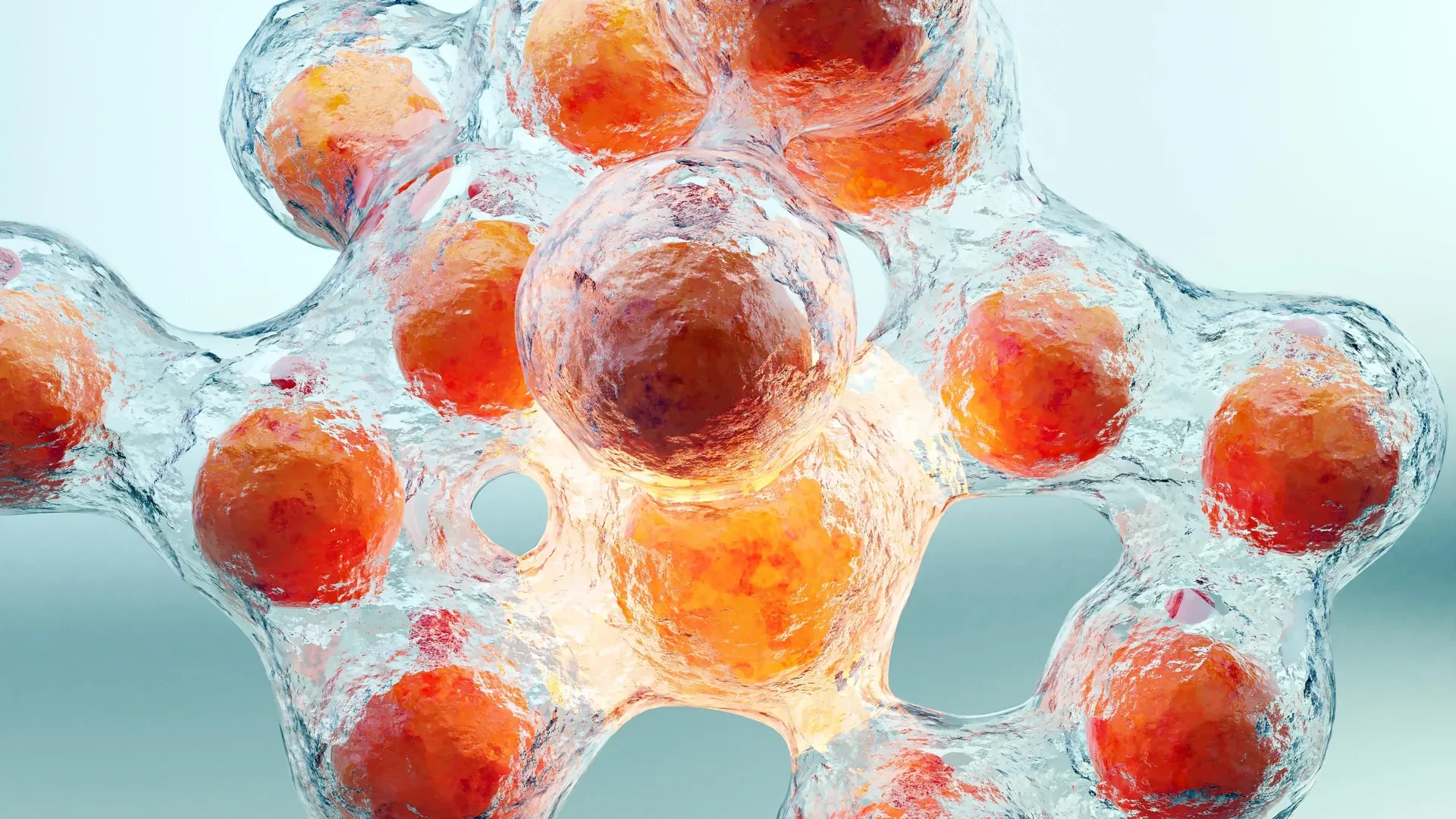
Adipose organoids are miniature, lab-grown three-dimensional tissue structures engineered to mimic the complex cellular architecture of real human fat tissue. By co-culturing primary human adipocytes with human immune cells—including eosinophils and macrophages—within a biocompatible extracellular matrix, researchers can observe real-time cellular crosstalk in an environment that faithfully mirrors human physiology.
The primary goals of this organoid engineering initiative include:
- Validating Subset Conservation: Determining whether the protective eosinophil subsets identified in female mice have direct functional counterparts in human female adipose tissue.
- Manipulating Intercellular Communication: Testing whether the application of human analogues to RELM alpha (such as human resistin or related signaling molecules) can restore anti-inflammatory macrophage polarization in organoid models subjected to inflammatory stress.
- Screening Sex-Specific Therapeutics: Utilizing organoids derived from both male and female human stem cell lines to screen immunomodulatory compounds, ensuring that potential anti-obesity drugs do not produce adverse sex-divergent side effects.
Towards Personalized Metabolic Medicine
The ultimate vision emerging from the UCR laboratory is the realization of truly personalized metabolic medicine. For decades, pharmacological interventions for obesity have focused primarily on appetite suppression (e.g., GLP-1 receptor agonists) or systemic metabolic rate enhancement. While undeniably revolutionary, these treatments often fail to address the underlying chronic tissue inflammation that drives insulin resistance, type 2 diabetes, and cardiovascular complications.
By shifting the therapeutic crosshairs toward the immunological microenvironment of fat tissue, future medicine may look very different. Imagine a class of precision biologics designed not to kill or broadly suppress immune cells, but to subtly reprogram specific eosinophil subsets—turning pro-inflammatory male-pattern immune profiles into protective, female-pattern cooperative networks.
Furthermore, because these mechanisms are inherently sex-differentiated, future clinical protocols will likely mandate sex-stratified dosing and pathway targeting, ensuring that treatments are tailor-made to harmonize with the unique immunological architecture of each patient.
As the UCR team continues its work with human adipose organoids, the scientific community moves one step closer to dissolving the boundary between immunology and metabolism—transforming our understanding of fat tissue from a passive liability into an actively managed, immunologically protected asset.
The study, titled "Single-cell heterogeneity of adipose eosinophils is shaped by sex and RELMα and linked to macrophage communication networks in obesity," was authored by Meera Nair, Adam Godzik, Rebecca E. Ruggiero-Ruff, Xinru Qiu, Lauren Vance, Yuxin He, and Djurdjica Coss. Funding was generously provided by the National Institutes of Health (NIH), the UCR School of Medicine, and the California Institute for Regenerative Medicine (CIRM).
