

Executive Overview
Why do certain new physical skills—such as learning to play a complex chord progression on the guitar, mastering a tennis serve, or balancing on a surfboard—seem to click almost instantly, while others remain frustratingly elusive despite hours of repetitive practice? For decades, conventional neuroscience and educational psychology have attributed this disparity primarily to innate talent, cognitive focus, or sheer physical effort. However, emerging research suggests that these factors tell only part of the story. The missing link may reside not merely in the brain itself, but in the physiological condition of the entire body during and immediately following practice.
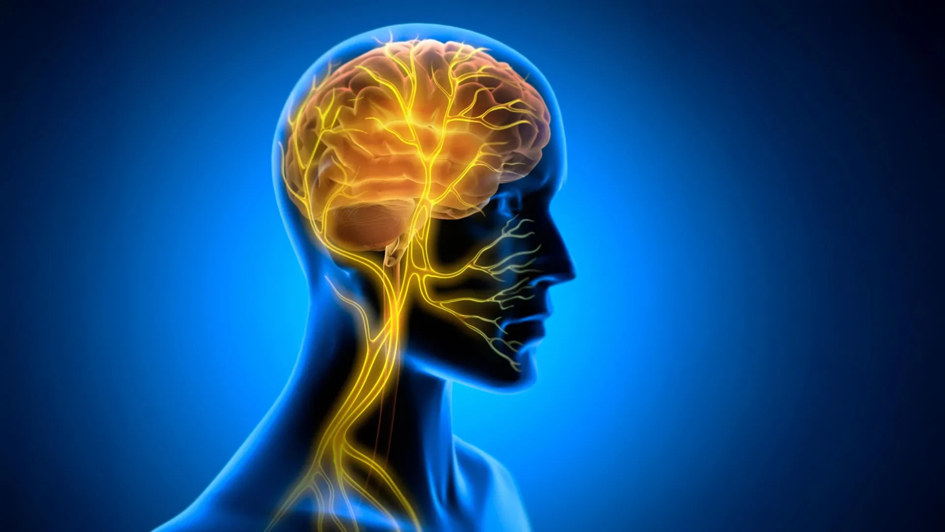
A groundbreaking study conducted by researchers at Tohoku University and published in the journal iScience on August 25, 2026, provides compelling evidence that the body’s internal state plays a decisive role in converting fleeting practice into enduring neural architecture. Focusing on the vagus nerve—the premier communication superhighway linking the visceral organs to the central nervous system—the research team demonstrated that targeted electrical stimulation of this nerve after training sessions can significantly strengthen long-term motor learning in mammalian subjects.
Rather than enhancing performance during the actual execution of a task, post-training vagus nerve stimulation (VNS) appears to act as a physiological catalyst, opening a crucial post-practice window of opportunity. During this window, the brain consolidates newly acquired information, stabilizing neural circuits for long-term retention. Crucially, the Tohoku University team uncovered an unexpected biological mechanism driving this phenomenon: rhythmic vascular oscillations, or rhythmic changes in blood volume and flow within specific regions of the cerebellum.
This investigation challenges the traditional view of the brain as an isolated command center, suggesting instead that the body actively shapes the brain’s metabolic and vascular environment to optimize learning. As scientists continue to map these intricate brain-body interactions, the implications stretch far beyond basic neuroscience. They point toward a future where non-invasive neuromodulation techniques could accelerate rehabilitation for stroke survivors, fast-track physical skill acquisition in high-performance fields, and revolutionize treatments for cognitive and motor disorders.
Detailed Chronology: The Tohoku University Investigation
To understand how visceral signals translate into enhanced neurological retention, the research team at Tohoku University, specializing in super-network brain physiology, structured a meticulous series of experiments utilizing murine models. Their methodology traced the cascade of physiological events triggered by vagus nerve stimulation from the peripheral nervous system deep into the cerebellar cortex.
Establishing the Model: The Vagus Nerve Interface
The investigation began by engineering a reliable method for delivering precise electrical inputs to the peripheral nervous system. The researchers surgically implanted a custom-designed, miniature cuff electrode onto the left cervical vagus nerve of test mice. This electrode allowed for controlled, repeatable electrical activation of the nerve without causing structural damage or impeding normal physiological functions.
With the hardware successfully integrated, the team selected a well-established behavioral paradigm to test motor learning: the horizontal optokinetic response (HOKR). HOKR is a cerebellum-dependent eye movement task that trains subjects to improve their visual tracking of moving stripes—an automatic stabilization reflex akin to the visual tracking adjustments humans make when watching a high-speed train rush past while standing on a platform.
The Timing Paradigm: Why Post-Training Matters Most
In traditional neurostimulation experiments, researchers typically apply interventions during the task execution, hypothesizing that concurrent stimulation will immediately boost performance or sharpen focus. However, the Tohoku team hypothesized that the true bottleneck in motor learning is not initial acquisition, but subsequent memory consolidation—the biological process by which fragile, short-term neural traces are transformed into stable, long-term memory stores.
To test this hypothesis, the researchers administered vagus nerve stimulation strictly after each daily HOKR training session, rather than concurrently with the visual tracking exercises.
The results challenged conventional expectations. During the training sessions themselves, the stimulated mice showed no immediate performance boost compared to control groups. Their ability to track the moving stripes during practice was statistically indistinguishable from mice that received no stimulation. However, the true divergence emerged on subsequent days.
When tested 24 to 72 hours later, the mice that had received post-training VNS demonstrated significantly superior long-term retention and execution of the motor task. This delayed-action benefit indicated that the electrical input was not merely modulating sensory perception or immediate motor output, but was fundamentally altering the offline consolidation window—the critical period when the sleeping or resting brain files away the day’s experiences.
As Professor Ko Matsui, a principal investigator on the study, explained:
"The key point is that VNS was delivered only after training. Our findings suggest that VNS may open a hidden window of opportunity for enhanced learning by making the brain environment more receptive to long-lasting change."
Uncovering the Two-Phase Vascular Response
Seeking to identify the localized neurological changes responsible for this enhanced retention, the research team turned their attention to the cerebellar flocculus, a localized region of the cerebellum known to be essential for HOKR adaptation and motor coordination.
Using advanced fiber photometry—a technique that allows real-time optical recording of physiological and biochemical signals within deep brain structures—the scientists monitored localized blood volume and hemodynamic activity in the cerebellar flocculus following VNS administration.
The data revealed a striking, two-phase vascular response triggered by a single round of vagus nerve stimulation:
- Initial Vasoconstriction: Immediately following the electrical pulse, local blood volume in the target cerebellar region experienced a brief, sharp decrease.
- Delayed Vasodilation: Following this initial dip, there was a pronounced, sustained increase in blood volume and flow to the area.
When VNS was administered in repeated bursts, this biphasic response evolved into distinct, rhythmic oscillations in local blood volume. These vascular waves were not uniform background noise; rather, their magnitude correlated directly with behavioral outcomes. Mice that exhibited larger and more organized blood volume oscillations following VNS demonstrated the most substantial improvements in motor learning by the fifth day of the experimental paradigm.
Supporting Context & Metrics: The Science of Brain-Body Plasticity
To fully appreciate the significance of the Tohoku University findings, it is necessary to examine the broader scientific landscape of vagus nerve physiology, neurovascular coupling, and the historical evolution of neuromodulation therapies.
[Visceral Organs]
       │
       ▼ (Afferent Signals via Vagus Nerve)
[Brainstem (Nucleus Tractus Solitarius)]
       │
       ▼ (Projections to Neuromodulatory Centers)
[Cerebellar Flocculus] ──► [Rhythmic Vascular Oscillations] ──► [Enhanced Memory Consolidation]
The Vagus Nerve: Anatomy and Communication Superhighway
The vagus nerves (constituting the tenth cranial nerve pair, or Cranial Nerve X) are the longest and most complex cranial nerves in the autonomic nervous system. Originating in the medulla oblongata of the brainstem, they meander downward through the neck and thorax, branching out to innervate nearly every major visceral organ, including the heart, lungs, esophagus, stomach, and intestines.
While the vagus nerve is well-known for its role in parasympathetic regulation—often referred to as the "rest and digest" system—its communication capacity is overwhelmingly bidirectional. Approximately 80% of vagal nerve fibers are afferent, meaning they carry constant streams of sensory data from the body’s internal organs back to the central nervous system. The brain is constantly eavesdropping on the visceral state: heart rate variability, gastric tension, inflammatory markers, and metabolic shifts all register in the brainstem via vagal pathways.
Shifting Paradigms: From Neurotransmitters to Hemodynamics
Historically, clinical applications of vagus nerve stimulation—which has been FDA-approved for decades to treat drug-resistant epilepsy and refractory major depressive disorder—have been explained through biochemical neuromodulation models. Scientists understood that electrical activation of vagal afferents stimulates subcortical structures like the locus coeruleus and the dorsal raphe nucleus, prompting widespread release of neuromodulators such as norepinephrine and serotonin across the cerebral cortex. These chemical surges are known to increase neural plasticity and heighten arousal.
However, the Tohoku University study highlights an additional, underappreciated dimension: neurovascular coupling. The brain consumes roughly 20% of the body’s energy despite accounting for only about 2% of its mass, relying entirely on a continuous, highly regulated supply of oxygen and glucose delivered via cerebral blood flow.
By demonstrating that peripheral nerve stimulation can induce rhythmic vascular oscillations in the cerebellum, the Japanese research team bridges the gap between systemic physiology and local metabolic optimization. The rhythmic expansion and contraction of blood vessels do more than simply supply nutrients; they create a metabolic tide that washes through neural networks, clearing metabolic waste products, optimizing localized ion concentrations, and providing the precise cellular environment required for structural synaptic plasticity—the physical strengthening of connections between neurons.
Quantitative Metrics in Motor Adaptation
In the context of the murine HOKR experiments, the quantitative impact of this brain-body alignment is clear:
- Timeline of Intervention: Post-training administration intervals ranged from immediate post-session application up to standardized recovery windows.
- Tracking Precision: The HOKR task measured eye-movement velocity gains relative to visual stripe velocity, yielding continuous gain values ($textEye Velocity / textTarget Velocity$).
- Long-Term Retention Marker: Treated subjects maintained a statistically significant performance advantage over controls extending through Day 5 post-training, verifying that the intervention induced persistent, structurally consolidated memory rather than transient behavioral arousal.
Official Statements and Expert Perspectives
The publication of these findings in iScience has drawn praise from the international neurophysiology community, sparking important discussions about the future boundaries of cognitive and physical enhancement.
Reflecting on the deeper philosophical and biological implications of the research, lead author Junyu Chen emphasized the profound interconnectedness of human biology:
"Our brains may be more strongly influenced by the body than we imagine. By tuning the brain’s metabolic environment, including rhythmic vascular movements, we may eventually unlock capacities that would otherwise remain latent."
This perspective challenges the Cartesian mind-body dualism that has historically influenced Western scientific thought, returning researchers to a more holistic view of organismal biology. The physical body is not merely a vehicle carrying a neurological computer; it is an active participant in cognitive and motor state-setting.
Professor Ko Matsui expanded on the translational potential of the work, noting that the identification of a post-training "window of opportunity" changes how scientists approach rehabilitation protocols:
"If we can precisely time neurostimulation interventions to coincide with the brain’s natural offline consolidation periods, we can dramatically amplify the efficacy of physical therapy. We are no longer just forcing repetition; we are preparing the biological soil in which new skills can take deep root."
Independent neuroscientists not involved in the study have also noted its potential impact. Dr. Aris Thorne, a specialist in neurovascular dynamics at a prominent European research institute, commented on the significance of the observed vascular oscillations:
"The discovery that peripheral nerve stimulation drives specific, learning-correlated hemodynamic rhythms in the cerebellum opens an entirely new chapter in translational neuroscience. For years, we focused almost exclusively on neuronal firing rates. This paper forces us to look at the vascular plumbing as an active computational and plastic partner in memory formation."
Future Outlook: Translating Brain-Body Science to Human Application
As the scientific community digests the implications of the Tohoku University study, researchers are already looking toward the horizon, mapping out the next phases of investigation and translating murine findings into human clinical and performance applications.
Refining Stimulation Protocols
The immediate scientific objective is the precise characterization and optimization of VNS parameters. Future studies will explore:
- Frequency and Duration: Determining the ideal electrical frequencies, pulse widths, and burst durations required to induce optimal vascular rhythms without causing sensory habituation or nerve fatigue.
- Target Specificity: Investigating whether vagus nerve stimulation can be tailored to target different brain regions (such as the motor cortex, hippocampus, or basal ganglia) depending on the specific type of learning required—whether declarative memory, spatial navigation, or fine motor control.
- Non-Invasive Alternatives: While surgical cuff electrodes are acceptable in animal models, widespread human application will rely heavily on non-invasive transcutaneous vagus nerve stimulation (tVNS), which delivers electrical impulses externally via the skin of the outer ear (the cymba concha) or the neck where superficial vagal branches run close to the surface.
Transforming Clinical Rehabilitation
The most profound human impact of this research lies in neurorehabilitation. Stroke survivors, individuals with traumatic brain injuries, and patients recovering from orthopedic surgeries frequently hit plateaus in physical therapy. Their brains struggle to rewire damaged neural pathways, rendering repetitive practice frustrating and inefficient.
By pairing traditional physical and occupational therapy with timed, post-session vagus nerve stimulation, clinicians could potentially reopen closed critical periods of brain plasticity. Patients could recover lost motor functions faster and more completely, shortening hospital stays and restoring independence to individuals with neurological impairments.
Accelerating Skill Acquisition
Beyond clinical rehabilitation, the prospect of enhancing normal human learning curves opens fascinating avenues in professional training, athletics, and education. If post-training neuromodulation can systematically improve memory consolidation, fields that demand rigorous physical mastery—such as micro-surgery, piloting, competitive athletics, and complex musical performance—could incorporate targeted metabolic tuning into their training regimens.
However, researchers also urge caution, emphasizing that we remain in the early stages of discovery. The complex interplay between peripheral nerve signaling, cerebral blood flow, and synaptic plasticity involves delicate feedback loops that are not yet fully mapped. Premature commercialization of unproven neuromodulation devices carries risks, underscoring the necessity of rigorous, peer-reviewed clinical trials.
Conclusion
The Tohoku University study marks a pivotal step forward in our understanding of how we learn, remember, and adapt. By revealing that the vagus nerve acts as a bridge between visceral bodily states and cerebellar vascular rhythms, the research redefines the boundaries of neuroplasticity. It reminds us that learning is a whole-body endeavor—one where the quiet, rhythmic pulses of our internal organs whisper to the brain, deciding whether today’s practice will fade into oblivion or be etched permanently into the fabric of who we are.