

Executive Overview
Alzheimer’s disease, the world’s most prevalent neurodegenerative disorder and the leading cause of dementia, has long eluded early-stage clinical intervention. For generations, medical science has approached the condition reactively, identifying the pathology only after patients exhibit unmistakable clinical symptoms such as memory loss, confusion, and behavioral changes. By the time these warning signs manifest, however, the underlying neurodegenerative process has often been compounding inside the brain for a decade or more, leaving behind an extensive and frequently irreversible trail of structural destruction.
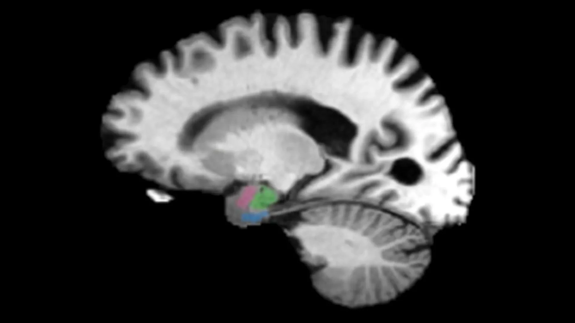
A paradigm-shifting study conducted by a multidisciplinary team of researchers at Johns Hopkins University threatens to rewrite this clinical narrative. By analyzing hundreds of advanced neuroimaging scans, the Johns Hopkins team has identified two specific subregions of the brain—the entorhinal cortex and the amygdala—that show detectable signs of tissue deterioration up to a decade before traditional memory symptoms emerge. Historically, clinical assessments of Alzheimer’s have heavily prioritized the hippocampus, viewing it as the primary historical anchor for early neurodegeneration. This new research reveals that tissue loss actually begins earlier and more aggressively in adjacent structures responsible for memory retrieval and emotional processing.
Published in NeuroImage Reports and featured prominently at Neuroscience 2026, the annual meeting of the Society for Neuroscience, these findings illuminate a critical window of opportunity. If clinicians can reliably identify individuals 5 to 10 years before cognitive decline becomes clinically apparent, therapeutic interventions can be administered when the brain is still resilient. Rather than attempting to rescue a heavily damaged organ, future medicine may possess the tools to intercept Alzheimer’s in its infancy, arresting its progression and preserving the cognitive identity of millions of patients worldwide.
Detailed Chronology: How the Breakthrough Unfolded
The path toward uncovering these ultra-early biomarkers required the synthesis of vast archival datasets, advanced neuroimaging analytics, and cutting-edge digital pathology. The investigation spanned several years, tracking biological transformations across diverse cohorts of patients.
Phase One: Harnessing Multicenter Datasets
To overcome the limitations of smaller, isolated sample sizes, the Johns Hopkins team turned to robust, standardized repositories. They examined hundreds of magnetic resonance imaging (MRI) and positron emission tomography (PET) scans sourced from two major, independent research frameworks: the Alzheimer’s Disease Neuroimaging Initiative (ADNI) and the Biomarkers of Cognitive Decline Among Normal Individuals (BIOCLAD) study.
These comprehensive datasets track individuals across a wide spectrum of cognitive health. The cohort included cognitively normal older adults, individuals experiencing mild cognitive impairment (MCI), and terminal patients whose brain tissues were available for post-mortem analysis. By analyzing longitudinal scans of these participants as they transitioned across different stages of health and disease, the researchers were able to trace the chronological footprint of structural brain changes.
Phase Two: Identifying the True Vanguard of Atrophy
Standard neurological evaluations for early-stage Alzheimer’s have traditionally focused on the hippocampus—a seahorse-shaped structure embedded deep within the temporal lobe that plays a central role in consolidating short-term memory into long-term storage. However, when the Johns Hopkins team mapped patterns of gray matter loss across the historical timeline of the disease, a different picture emerged.
The data revealed a consistent, highly predictable anatomical pattern. While the hippocampus certainly deteriorates, two neighboring structures—the entorhinal cortex and the amygdala—undergo relative tissue shrinkage earlier in the disease timeline.
- The Entorhinal Cortex: Serving as a primary gateway between the hippocampus and the neocortex, this region is vital for navigating spatial environments and retrieving previously stored episodic memories.
- The Amygdala: Positioned adjacent to the hippocampus, this almond-shaped cluster of nuclei governs emotional processing, fear conditioning, and the formation of emotionally charged memories.
The early degradation of these two interconnected regions provides a revised chronological blueprint for how Alzheimer’s establishes its stronghold within the human central nervous system.
Phase Three: Connecting Atrophy to the Tau Protein
Recognizing anatomical shrinkage is only half the battle; medical science must also understand the biochemical engine driving that physical deterioration. To bridge this gap, the researchers combined their structural MRI findings with PET imaging and digital pathology techniques to track the presence of tau—a hallmark protein implicated in Alzheimer’s pathology.
Under homeostatic conditions, healthy tau proteins stabilize microtubules, which form the internal skeletal infrastructure of neurons and facilitate the transport of vital nutrients. In the pathological cascade of Alzheimer’s, however, tau undergoes hyperphosphorylation, detaches from microtubules, and misfolds into insoluble neurofibrillary tangles. These tangles choke the internal machinery of neurons, leading to cell death and the subsequent loss of gray matter.
By cross-referencing their imaging data, the Johns Hopkins team discovered a striking spatial overlap: the regions exhibiting early gray matter atrophy (the entorhinal cortex and the amygdala) mapped directly onto the zones showing early tau-related accumulation. This biochemical correlation bridges the macroscopic observation of brain shrinkage with the microscopic reality of cellular protein pathology.
Supporting Context & Metrics: The Scale of the Challenge
To fully appreciate the significance of the Johns Hopkins discovery, one must examine the broader epidemiological and biological landscape of Alzheimer’s disease.
The Global Burden of Neurodegeneration
Alzheimer’s disease is not merely a natural consequence of aging; it is a progressive, fatal neurodegenerative disorder. According to global health metrics, tens of millions of individuals worldwide currently live with dementia, with Alzheimer’s accounting for roughly 60% to 80% of all cases. As global life expectancies rise, the prevalence of the disease is projected to double every two decades, threatening to overwhelm healthcare infrastructures, caregivers, and global economies.
Despite decades of intensive pharmacological research, treatments capable of reversing Alzheimer’s do not yet exist. Recent therapeutic breakthroughs—such as monoclonal antibody treatments designed to clear amyloid-beta plaques from the brain—have demonstrated an ability to modestly slow cognitive decline, but only when administered in the earliest stages of the disease. Once widespread neuronal death has occurred, clearing proteins cannot resurrect dead brain cells. This economic and clinical reality underscores why identifying pre-symptomatic biomarkers is the holy grail of modern neurology.
The Silent Decades: A Biology of Attrition
Pathological studies have long indicated that the biological machinery of Alzheimer’s operates in silence for years before a patient forgets where they left their keys or struggles to find a word. This "silent phase" is characterized by two primary biochemical adversaries:
- Amyloid-beta plaques: Clumps of protein fragments that accumulate outside neurons, disrupting intercellular communication.
- Neurofibrillary tau tangles: Intracellular aggregations that destroy the structural integrity of the neuron itself.
For years, researchers debated the exact timeline of these accumulations. The new Johns Hopkins study provides quantitative weight to the hypothesis that structural atrophy in specific limbic subregions tracks alongside these biochemical shifts up to a decade before clinical diagnosis. By treating this 5-to-10-year pre-symptomatic window as a therapeutic target, medicine shifts its posture from palliative management to preventive intervention.
Official Statements: Perspectives from the Research Team
The gravity of the findings has been articulated by the study’s principal architects, who emphasize both the immediate promise and the long-term clinical implications of their work.
Dr. Michael Miller, a biomedical engineer and senior author of the study, emphasized the profound shift in timing that these biomarkers represent:
"We have found that there are early changes in the brain manifesting the clinical biomarkers of atrophy as much as a decade before there are clinical symptoms. It’s at least a 5-to-10-year play where your brain is changing. Imagine if we could start to treat the neurodegenerative changes at the 2, 3 or 4-year point rather than after 15 years when the changes are manifesting in memory symptoms. That’s the great promise here for your loved ones who are at risk."
Dr. Marilyn Albert, co-author and director of the Johns Hopkins Alzheimer’s Disease Research Center, highlighted the ultimate clinical objective of the research initiative:
"Our goal is to be able to treat individuals before the symptoms of Alzheimer’s disease develop so that they may be delayed or even stopped."
Addressing the practical utility of the discovery for risk stratification, co-author and neuropathologist Dr. Meaghan Morris explained how these findings could transform preventive care:
"As biomarkers and treatments become increasingly available, knowing who is going to have cognitive decline is going to be really important. This could really play into better risk stratification for identifying who is going to have cognitive decline in the near future—and treating people earlier and earlier to hopefully slow the course of the disease."
Looking toward the future of cellular mapping, Dr. Miller outlined the next phase of the team’s ongoing investigation:
"Our future work is to better show the molecular neighborhoods associated with these Tau protein changes as localized by these clinical biomarkers of atrophy. Once we know more about the molecular neighborhoods, we can develop therapies."
Future Outlook: Translating Discovery into Clinical Practice
While the identification of early tissue loss in the entorhinal cortex and amygdala marks a watershed moment in neuroimaging research, significant hurdles remain before these insights translate into routine clinical diagnostics.
From Research Tool to Clinical Diagnostic
At present, the detection of subtle volumetric changes in deep-brain subregions requires sophisticated neuroimaging software, high-field MRI scanners, and expert radiological analysis. For this discovery to impact the general population, these analytical pipelines must be streamlined, standardized, and made economically viable for widespread clinical use.
As imaging hardware becomes increasingly sensitive and less expensive, researchers envision a future where routine brain scans—perhaps incorporated into mid-life health screenings for individuals with a strong family history of dementia—could evaluate the health of the entorhinal cortex and amygdala as early as a person’s 30s or 40s. Just as routine blood panels screen for cholesterol levels and cardiovascular risk decades before a heart attack, future neurological check-ups could profile structural brain integrity to assess neurodegenerative risk.
Decoding the "Molecular Neighborhoods"
The Johns Hopkins team is already looking beyond macroscopic structural scans. Their next investigative frontier involves dissecting the precise molecular environments—the "molecular neighborhoods"—that surround tau protein accumulations within the entorhinal cortex and amygdala.
By understanding the micro-scale cellular dynamics that trigger tau hyperphosphorylation and subsequent gray matter atrophy, pharmaceutical developers can design targeted molecular therapies. These interventions would aim to interrupt the disease process at its cellular roots, neutralizing toxic proteins and halting tissue shrinkage before it can cascade into the hippocampus and wider cerebral cortex.
A Beacon of Hope for Future Generations
The war against Alzheimer’s disease has been characterized by high-profile clinical trial failures and heartbreaking losses for patients and their families. However, the identification of ultra-early structural biomarkers changes the strategic calculus of neurodegenerative medicine. By illuminating the silent, decade-long runway that precedes clinical symptoms, Johns Hopkins researchers have provided science with a vital map. If leveraged correctly, this map will guide humanity toward an era where Alzheimer’s disease is no longer an inevitable tragedy of aging, but a preventable, treatable condition.
