

Executive Overview
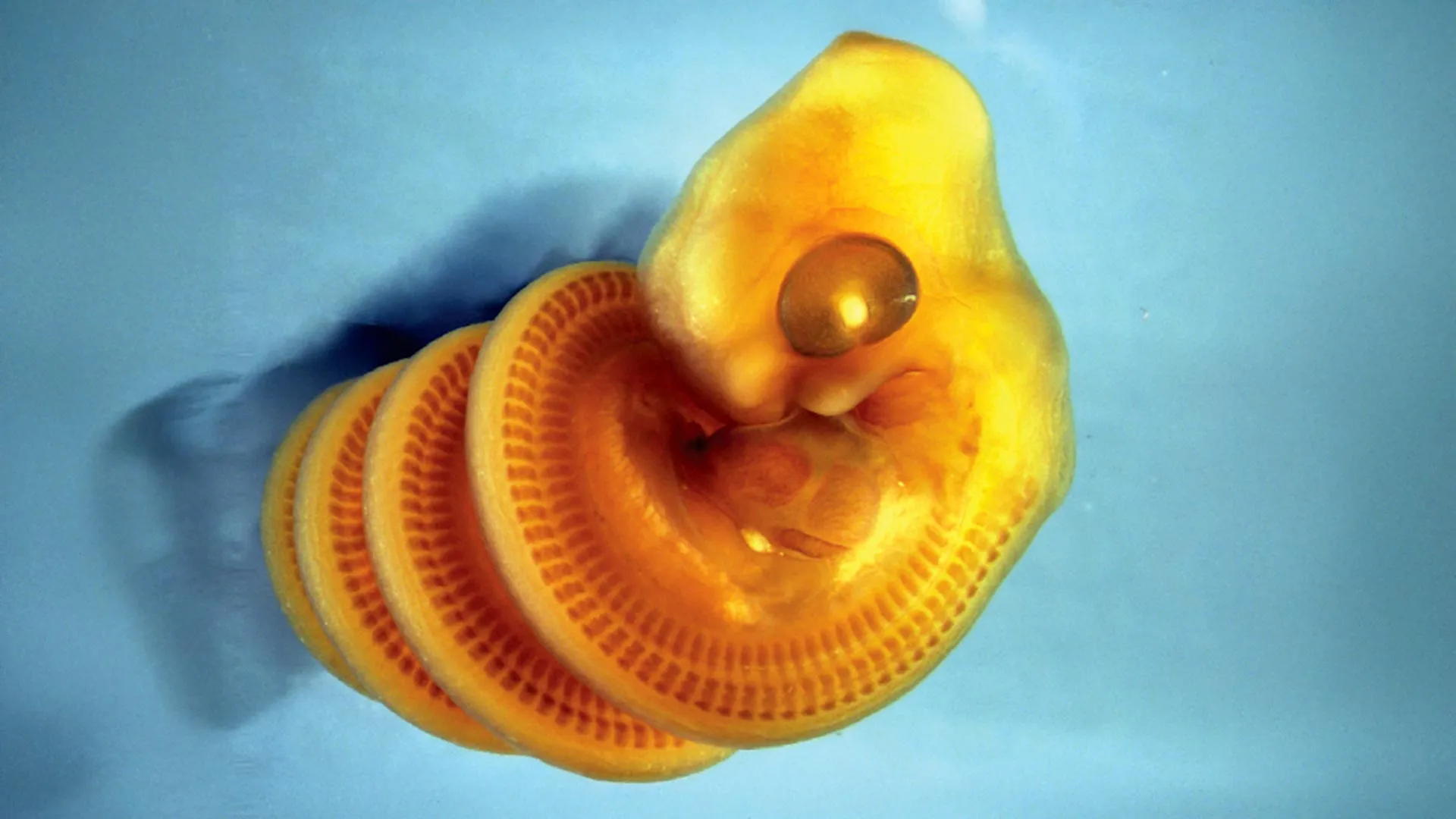
For centuries, the evolutionary trajectory of snakes has captivated and confounded naturalists. Characterized by an extraordinary elongation of the vertebrate body plan—featuring hundreds of vertebrae, the complete loss of limbs, and the rearrangement of internal organs—serpents represent one of nature’s most radical anatomical departures. Yet, long before these creatures slither into the light of day, a profound physical transformation occurs within the confines of the egg: developing snake embryos curl into tight, mathematically precise spirals.
Until recently, the driving mechanism behind this peculiar pre-hatching posture remained an enduring biological enigma. Was it an active muscular choice? A genetic predisposition? Or something far more fundamental?
An international research team, spearheaded by scientists in Canada and published in the journal Current Biology, has finally unraveled this mystery. By analyzing more than 900 embryos across 39 distinct snake and limbless squamate species, the researchers discovered that this coiling behavior is not a muscular feat, but rather a brilliant mechanical solution to an architectural crisis. As a snake embryo grows at an explosive rate, its digestive tract lags behind, acting as a physical tether. This growth mismatch forces the rapidly lengthening body to buckle, twist, and naturally curl into a uniform, right-handed spiral.
This discovery does more than merely explain a peculiar developmental quirk of reptiles; it introduces a compelling new model for understanding how asymmetrical and spiral structures manifest across the animal kingdom. Born from the unprecedented isolation of the 2020 COVID-19 lockdowns, this study stands as a testament to the power of observational biology, proving that monumental insights can sometimes be unearthed not through high-tech genetic sequencing, but by simply pausing to look closely at the physical forces shaping life.
Detailed Chronology: From Lockdown Curio to Biological Breakthrough
The Genesis of an Idea: 2020 Isolation
The roots of this scientific breakthrough trace back to the spring of 2020. As the COVID-19 pandemic brought global society to a sudden, grinding halt, Dr. Tetsuto Miyashita, an evolutionary biologist at the Canadian Museum of Nature, found himself working remotely from home. Like countless researchers worldwide, Miyashita was temporarily locked out of laboratories, field sites, and physical museum collections.
Faced with these unprecedented constraints, Miyashita set out to formulate a research question that his students could investigate entirely through remote digital resources—a project that required no wet labs, no live animal handling, and no physical museum access.
"Then the lightbulb turned on," Miyashita recalls. "I had inherited from my PhD advisor this fascination with asymmetries in animal forms. So every time I saw images of snake embryos in papers, I wondered whether they are right- or left-handed in their coiling."
This simple, whimsical curiosity regarding the "handedness" of snake embryos became the foundational seed of a multi-institutional study. Miyashita enlisted Alexandra Weber—then a student at Carleton University (now a graduate student in zoology at the University of British Columbia)—alongside two undergraduate researchers at the University of Ottawa, tasking them with an exhaustive digital scavenger hunt.
Combing the Archives: The 900-Embryo Dataset
The team scoured published scientific literature, historical archives, and digital museum databases, harvesting photographs and illustrations of developing snake embryos. The scope of the collection quickly expanded far beyond initial expectations.
Ultimately, the researchers compiled a statistically robust sample comprising more than 900 individual embryos representing 39 distinct species of snakes and other limbless squamates (lizards that have evolutionarily lost their limbs).
When the team analyzed the compiled imagery, a striking and undeniable pattern emerged. During the earliest developmental phases—the first several weeks following egg-laying—every single observed embryo exhibited the exact same spatial orientation. They were exclusively dextral, meaning they coiled strictly to the right when viewed along the axis from head to tail.
"At these stages, the embryos don’t have muscles to move with, so different forces are making them coil right-handed," explains Alexandra Weber, lead author of the study. "But we didn’t know what’s making them do that."
Because muscular movement was entirely out of the equation during these early windows, the research team realized that the answer must lie in pure physics and developmental biomechanics. Something physical was forcing the embryos into this rigid, predictable geometry.
The CT Scan Revelation
To peer inside the microscopic anatomy of these embryos with unprecedented clarity, the team turned to Dr. Raul Diaz, a collaborator at California State University, Los Angeles. Utilizing advanced computed tomography (CT) imaging, Diaz began scanning snake embryos to map their internal structures in three dimensions.
The resultant scans exposed a hidden anatomical arrangement that had previously gone unnoticed by generations of comparative anatomists.
"Raul’s CT scan of a snake embryo revealed a structure we had never seen before—it was a pillar of gut stretching through the spiral of the coiling body," Miyashita explains. "There’s an intestine detached from the rest of the body, surrounded by tendrils of blood vessels from the yolk."
This anatomical revelation provided the missing mechanical puzzle piece. Snake embryos are evolutionarily programmed to lengthen at an astonishingly rapid pace to achieve their characteristic, elongated body plans. However, the embryonic gut does not expand at this same breakneck speed. This stark mismatch in growth rates generates immense mechanical stress.
Because the slow-growing gut remains anchored, it acts as a physical tether against the aggressively expanding body wall. The body has nowhere to go; as it continues to elongate against this internal anchor, it buckles.
Furthermore, the physical positioning of the yolk provides the directional bias. The yolk consistently rests on the left side of the developing snake embryo. Consequently, when the buckling force is released, it is directed toward the opposite side, inevitably forcing the embryo to twist and coil exclusively to the right.
The Shift to Freedom: Later-Stage Development
Crucially, this right-handed lock is not permanent. As incubation progresses, the dynamics within the egg shift dramatically. The yolk gradually depletes and shrinks, creating spatial relief within the eggshell. Simultaneously, the embryonic muscles mature and strengthen, granting the young snakes the agency of independent motion.
"Some remain in right-handed coils, but some recoil to the left side," Weber notes. "So half of these near-hatching embryos are right-handed and the other half left-handed."
By the time the snakes are fully prepared to pip their shells and emerge into the world, the strict directional bias observed in early development has dissolved into a balanced, random distribution of left- and right-handed coils. This confirmed that the initial coiling is an entirely passive, mechanically driven phenomenon rather than a behavioral choice or a lifelong anatomical trait.
Supporting Context & Metrics
To fully appreciate the scope and significance of this study, it is helpful to examine the quantitative framework and the broader biological context surrounding serpentine evolution.
| Research Parameter | Metric / Detail |
|---|---|
| Total Embryos Examined | > 900 individual specimens |
| Species Diversity | 39 distinct snake and limbless squamate species |
| Initial Coiling Direction | 100% dextral (right-handed) during early developmental stages |
| Final Coiling Distribution | 50% right-handed, 50% left-handed near-hatching stage |
| Key Anatomical Constraint | Mismatch in growth rate between the rapidly lengthening body and the slow-growing gut (intestine) |
| Primary Driver of Asymmetry | The yolk sac, which consistently rests on the left side of the embryo, forcing initial buckling to the right |
| Primary Methodology | Digital archival search, comparative anatomical analysis, and high-resolution CT imaging |
The Evolutionary Context of Serpentine Elongation
Snakes diverged from lizard ancestors over 100 million years ago, embarking on an evolutionary experiment defined by extreme body elongation. While most vertebrates maintain a relatively compact skeletal ratio, snakes possess anywhere from 130 to over 400 vertebrae. Accommodating this staggering length within a confined, spherical or oval eggshell presents an immense spatial challenge.
Nature’s solution is embryonic coiling. Without this tight, helical packing strategy, an elongated vertebrate embryo would be structurally incapable of fitting inside a standard amniotic egg. By identifying the mechanical interplay between a tethering gut and an expanding body wall, this study sheds light on how physical constraints directly dictate developmental morphology in extreme phenotypes.
Official Statements and Expert Perspectives
The collaborative nature of this international study brought together diverse academic disciplines—ranging from paleontology and evolutionary biology to zoology and bioimaging. Here is what the leading researchers have to say about their findings:
Dr. Tetsuto Miyashita (Senior Author & Evolutionary Biologist, Canadian Museum of Nature):
"There is a touch of mystery to spirals, and we are only beginning to understand how these shapes are produced in animals, such as our looping intestine, snail shells, and now these beautifully coiled snake embryos… These puzzles beckon our curiosity. After all, spiral forms in nature have inspired human creations ranging from rotini pasta, to a barber’s pole or even portrayals of the biblical ‘Tower of Babel’."Alexandra Weber (Lead Author & Zoology Graduate Student, University of British Columbia):
"At these stages, the embryos don’t have muscles to move with, so different forces are making them coil right-handed… But we didn’t know what’s making them do that. This all started out with a curiosity to see if snakes are ‘handed’. It was exciting to follow it to deep insights about their evolution."Reflecting on the Simplicity of the Discovery:
"Scientists have long been fascinated with how and why snakes evolved their strange body form. To answer that question, they tended to take a deep dive into sophisticated genetic research, looking at Hox genes, enhancers, and so on… These are key discoveries. But here, out of the COVID lockdown, we uncovered a snake’s secret with a startlingly simple approach—just scroll through an album of snake embryos and record which way they are coiled, and take a good look at their anatomy." — Dr. Tetsuto Miyashita
Future Outlook: A New Paradigm for Biological Spirals
Beyond resolving a niche question in reptilian embryology, the implications of this research extend far into broader developmental biology and biophysics. Spirals and helical forms are ubiquitous in the natural world—manifesting in DNA double helices, cardiac muscle fiber architecture, cochlear structures in the inner ear, gastrointestinal loops, and the intricate shell geometries of mollusks.
Historically, explaining these complex geometries has required invoking intricate genetic regulatory networks or complex biochemical gradients. However, the Canadian-led team’s findings suggest that simple, elegant mechanical principles—such as growth rate mismatches and physical tethering by internal organs—may play a vastly underappreciated role in shaping embryonic anatomy.
Expanding the Model
Buoyed by their success with snake embryos, the research team is actively exploring how this mechanical buckling model can be adapted to study other spiral-shaped biological structures. By integrating developmental biology with biomechanical modeling, scientists hope to determine whether similar growth-constraint mechanics govern organ looping in mammals, plant tendril coiling, or the asymmetrical positioning of internal organs (situs inversus) in vertebrates.
Furthermore, this study serves as an inspiring methodological reminder for the scientific community. In an era dominated by high-throughput sequencing, CRISPR-Cas9 knockouts, and big-data bioinformatics, the breakthrough demonstrates that deep, fundamental insights are still waiting to be discovered through basic observational inquiry, cross-institutional collaboration, and a childlike sense of curiosity.
As researchers continue to refine this new model of embryonic biophysics, the humble snake embryo—neatly curled in its right-handed helix—has proven to be an unexpected masterclass in how physical forces write the blueprints of life.