

Executive Overview
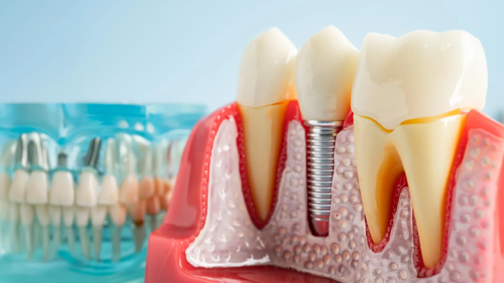
For tens of millions of people worldwide, dental implants have represented a revolutionary leap forward in restorative medicine. Offering a permanent, fixed alternative to removable dentures, implants restore both functionality and aesthetics, allowing patients to eat, speak, and smile with confidence. Yet, beneath this clinical success story lies a persistent and painful complication: peri-implantitis. Estimated to affect between 10% and 20% of all implant recipients, this aggressive inflammatory condition progressively destroys the jawbone anchoring the prosthetic, often leading to implant failure.
For years, the medical and dental communities have been baffled by a frustrating clinical reality. While peri-implantitis begins with the same oral bacteria responsible for standard gum disease (periodontitis)—a condition easily managed with professional cleanings and antibiotics—infections surrounding dental implants stubbornly resist conventional therapies. Traditional antibiotic regimens fail in more than half of all cases, leaving clinicians with few options outside of surgical intervention or implant removal.
Now, groundbreaking research published in PNAS Nexus by a team at the Rutgers School of Dental Medicine has unmasked the cellular mechanism driving this phenomenon. The study reveals that oral bacteria do not act alone; instead, they create acidic biofilms that slowly corrode the titanium body of the implant itself. This corrosion sheds billions of microscopic metal particles into the surrounding gum tissue.
Far from being inert, these titanium fragments act as molecular " Trojan horses." Coated in bacterial toxins, they trick the body’s primary infection-fighting white blood cells—macrophages—into an endless, destructive state of hyper-inflammation. While trapped in this futile battle against un-digestible metal, the immune system abandons its fight against the actual bacteria, creating a cascading failure of host defenses. Crucially, the Rutgers team has identified the specific cellular calcium channel responsible for this cascade, pointing the way toward the world’s first targeted drug therapies for peri-implantitis. With healthcare systems worldwide absorbing over $1 billion annually in complication costs, this discovery promises to fundamentally alter how dental implant diseases are prevented, managed, and treated.
Detailed Chronology of Discovery
Decades of Frustration: The Antibiotic Paradox
To understand the significance of the Rutgers breakthrough, one must look back at two decades of clinical confusion. When dental implants surged in popularity during the late 20th century, clinicians assumed that infections surrounding them would behave identically to infections around natural teeth. Both environments harbor analogous microbial communities, dominated by gram-negative anaerobic bacteria.
However, clinical outcomes told a drastically different story. While traditional periodontitis reliably recedes when treated with mechanical debridement and systemic or localized antibiotics, peri-implantitis proved notoriously refractory. Standard antibiotic therapies achieved clinical success in fewer than 40% to 50% of implant cases. Even aggressive pharmacological interventions often failed to halt the underlying degradation of the alveolar bone.
For nearly twenty years, researchers fixated almost exclusively on the microbial side of the equation, searching for hyper-virulent bacterial strains or unique biofilm structures. It was only recently that senior author Georgios Kotsakis and his laboratory shifted the investigative paradigm, asking a radically different question: What role does the implant material itself play in driving this chronic inflammation?
The Rutgers Investigation: Tissue, Lab, and Animal Models
To test the hypothesis that the implant material contributes to the disease pathology, the research team at Rutgers School of Dental Medicine mounted a multi-tiered investigation. They combined clinical tissue analysis with advanced cellular biology and in vivo mammalian testing.
- Human Tissue Samples: The researchers first analyzed diseased gum and bone tissues retrieved from human patients suffering from advanced peri-implantitis, identifying the presence of microscopic wear debris.
- In Vitro Cellular Studies: Human macrophages were isolated and cultured in laboratory environments, then exposed to varying concentrations of titanium particles and bacterial lipopolysaccharides to observe real-time behavioral changes.
- Genetically Engineered Animal Models: To prove causation rather than mere correlation, the team utilized genetically modified mice engineered to lack a specific calcium channel protein. These murine models were subjected to the same pathological conditions to observe whether blocking the pathway could alter disease progression.
Pinpointing the Culprit: TRPC1 and Cellular Hijacking
Through this rigorous methodology, the Rutgers team mapped out the complete pathological sequence:
- Acidic Biofilm Formation: Bacteria accumulating on the rough surfaces of the titanium implant generate localized acidic microenvironments.
- Corrosion and Shedding: Over time, this acid breaks down the titanium oxide passivation layer of the implant, shedding billions of microscopic particles—each significantly smaller than a red blood cell. Mechanical cleaning instruments can also dislodge these fragments.
- Toxin Coating: Once free in the tissue, these titanium fragments attract lipopolysaccharides (LPS), a potent bacterial toxin. This creates a deceptive composite structure that mimics an unusually large, indestructible bacterium.
- Macrophage Entrapment: Macrophages attempt to phagocytose (engulf) the toxin-coated particles. Because titanium cannot be enzymatically broken down, the macrophages become chronically activated, generating massive quantities of inflammatory cytokines—specifically interleukin-1 beta (IL-1β), a protein heavily implicated in destructive autoimmune and inflammatory disorders like rheumatoid arthritis.
- The Immunity Trade-Off: Saturated by their futile attempt to digest metal, the macrophages lose their functional capacity to target actual bacteria. Laboratory assays demonstrated that titanium-exposed macrophages engulfed fewer than half as many bacteria as healthy control cells.
Finally, the team tracked this hyperactivity down to a specific molecular gateway: TRPC1, a pore-forming calcium channel nestled within the macrophage cell membrane. When TRPC1 is stimulated by the titanium-LPS complex, it floods the cell with calcium ions, locking the immune response into its hyper-inflammatory loop.
Supporting Context & Metrics
The clinical and economic footprint of dental implant complications is staggering. As surgical techniques have improved, millions of patients have opted for implants over bridges and dentures. However, the long-term maintenance burden has grown commensurately.
Key Metrics in Implant Dentistry
- 10% to 20%: The estimated percentage of dental implant recipients who ultimately develop peri-implantitis.
- $1 Billion+: The annual financial burden imposed on global healthcare systems by peri-implantitis treatments, revision surgeries, and implant removals.
- < 50%: The clinical success rate of traditional antibiotic therapies when applied to peri-implantitis, compared to high efficacy rates in natural tooth periodontitis.
- Sub-cellular Scale: The size of the shed titanium particles, each measuring fractions of a micron—smaller than a red blood cell, allowing them to deeply infiltrate surrounding soft and hard tissues.
The Mechanical Complication Factor
Beyond natural corrosion driven by bacterial biofilms, the study highlighted iatrogenic risks—specifically, how dental professionals clean implants. For decades, standard dental prophylaxis involved metal scalers designed to scrape hardened calculus (tartar) off natural enamel.
When applied to softer titanium implant surfaces, these metal instruments gouge, scratch, and pit the metal, inadvertently shedding thousands of micro-particles into the gingival pocket. Recognizing this hazard, modern clinical standards have shifted toward non-abrasive instruments—such as specialized plastic, resin, or titanium-coated ultrasonic tips, along with air-polishing devices using amino acid powders—to minimize surface disruption and subsequent particle shedding.
Official Statements
The findings have sent ripples through the dental research and clinical communities, offering a cohesive explanation for a medical mystery that has plagued practitioners for decades.
"For the first time, we show why all the antibiotic treatments that work around teeth do not work around implants. Now that we know the cause, we can start developing therapeutics."
— Georgios Kotsakis, DDS, MS, Senior Author of the study and Assistant Dean for Clinical Research at the Rutgers School of Dental Medicine.
Kotsakis emphasized the deceitful nature of the biological mechanism:
"These particles are little magnets that attract the bacterial toxin, and they hijack the immune system, preventing it from clearing bacteria. You have a perfect storm that defies antibiotics."
Reflecting on the therapeutic implications of the animal trials—where disabling the TRPC1 calcium channel successfully halted bone destruction and reduced abscess formation—the research team noted that shifting the therapeutic paradigm from killing bacteria to modulating the host immune response represents a watershed moment for oral medicine.
Future Outlook: Therapeutics and Prevention
The identification of TRPC1 as the critical molecular switch in peri-implantitis opens an entirely new frontier in dental pharmacology. Rather than relying on broad-spectrum antibiotics that fail to penetrate the immune paralysis caused by titanium debris, future treatments may involve localized or systemic inhibitors designed to block the TRPC1 calcium channel.
The Road to Clinical Trials
With critical funding secured from the National Institutes of Health (NIH), Dr. Kotsakis and his colleagues at Rutgers are actively evaluating candidate pharmacological agents capable of safely inhibiting the TRPC1 pathway in human cells. If these preclinical evaluations prove successful, human clinical trials could eventually yield the world’s first FDA-approved host-modulation drugs specifically indicated for peri-implantitis.
Preventive Paradigm Shifts
While pharmacological treatments wind their way through the development pipeline, the study reinforces the absolute necessity of rigorous preventative maintenance:
- Advanced Prophylactic Techniques: Dental practitioners must universally adopt non-abrasive cleaning technologies (such as specialized air-polishing and carbon-fiber or resin scalers) to prevent iatrogenic titanium shedding.
- Early Interventions: Because peri-implantitis is insidious and bone loss can occur rapidly once the inflammatory cascade begins, routine radiographic monitoring and early detection of sub-gingival biofilm accumulation remain paramount.
- Biomaterial Engineering: In the long term, this research provides vital feedback to implant manufacturers. Insights into how acidic biofilms degrade titanium surfaces will undoubtedly drive the development of next-generation implant coatings, alloy compositions, and surface topographies engineered to resist both bacterial colonization and corrosion.
By illuminating the hidden interplay between metal, microbes, and mammalian immunity, the Rutgers study transforms peri-implantitis from an intractable clinical mystery into a solvable engineering and pharmacological challenge, ensuring that dental implants can fulfill their promise of lifelong restorative health.