

Executive Overview
In the rapidly evolving frontier of biohybrid robotics, a paradigm shift is underway. For decades, roboticists have relied on traditional, energy-dense hardware—heavy metals, rigid plastics, complex gearboxes, and bulky electric motors—to achieve mechanical motion. However, a pioneering team of researchers at the Massachusetts Institute of Technology (MIT) has shattered these conventions. In a study recently published in the prestigious journal Advanced Functional Materials, the MIT team demonstrated that an exceptionally thin, two-dimensional layer of living muscle tissue can generate sufficient force to propel a fully functional, untethered swimming robot through water.
Measuring roughly the length and width of a standard stick of gum, this ultra-thin aquatic device bypasses the need for conventional mechanical actuators. Instead, it relies on genetically modified skeletal muscle cells grown atop a flexible, highly optimized gel framework. By simply shining light onto specific sections of the robot, the researchers can trigger coordinated muscle contractions that flap the device’s dual fins, steering it smoothly through complex underwater mazes.
Led by Ritu Raman, associate professor of mechanical engineering at MIT, this breakthrough represents the first successful demonstration of a micro-scale biohybrid robot powered by a two-dimensional living muscle architecture. While previous iterations of biohybrid machines required massive, three-dimensional blocks of lab-grown tissue comprising millions of cells, this new design achieves superior movement efficiency with a fraction of the biological material.
The implications of this research extend far beyond aquatic locomotion. By harnessing the unique properties of living tissue—such as inherent compliance, environmental responsiveness, and autonomous self-healing capabilities—this technology paves the way for a new generation of soft, adaptable robots. These systems could soon be deployed in delicate, unpredictable ecosystems where traditional hard-bodied hardware would cause ecological damage or fail to navigate tightly constrained spaces.
Detailed Chronology
The creation of MIT’s muscle-powered swimmer did not happen overnight; it is the culmination of years of iterative biomaterials engineering, fundamental cell biology research, and mechanical optimization.
Phase I: The Artificial Iris and the 2D Muscle Foundation
The foundational concepts behind the new swimming robot originated in Raman’s laboratory last year, when the research team set out to engineer an artificial muscle disk inspired by the mechanics of the human eye’s iris.
To achieve this, the engineers fabricated a soft gel disk patterned with microscopic grooves radiating outward from its center and intersecting to form concentric circles. Upon this patterned surface, they seeded living muscle cells. As the cells proliferated, they naturally followed the topological cues of the micro-channels, organizing themselves into a continuous, ultra-thin biological layer.
When exposed to light, the engineered tissue contracted multi-directionally, forcing the underlying gel disk to stretch and compress in a manner mimicking how a human pupil expands and narrows in response to light levels. While this artificial iris successfully proved that scientists could grow exceptionally thin, responsive muscle layers capable of complex movements, its practical applications in robotics were severely limited. The total displacement of the tissue was restricted to roughly 100 microns—a microscopic movement that, from a robotic propulsion standpoint, was far too small to achieve meaningful work.
Phase II: Overcoming Material Limitations
Buoyed by the success of the artificial iris, the research team established a much more ambitious objective: dramatically scaling up the force output and physical displacement of the muscle cells to propel an untethered robot through fluid media.
This ambition immediately exposed a critical limitation in their previous methodology. The artificial iris had relied on fibrin, an exceptionally soft biological gel, as its underlying substrate. However, as the researchers quickly discovered, fibrin was fundamentally unsuited for high-force applications. Under the intense mechanical tension exerted by contracting muscle fibers, the soft fibrin matrix rapidly shrank and degraded, severely dampening the force transferred to the surrounding environment.
Recognizing that they needed a more robust foundation, the engineers systematically investigated three foundational parameters of the gel substrate:
- Chemical composition: Moving away from soft fibrin to more resilient biomaterials.
- Substrate stiffness: Determining the optimal modulus of elasticity required to resist excessive contraction while still supporting cellular attachment.
- Dimensions and geometry: Refining the micro-scale surface patterns used to guide cellular alignment and tissue fusion.
Phase III: Optimizing the Gel Substrate and Cellular Alignment
To identify the ideal structural foundation, the MIT team tested a variety of formulations, stiffness levels, and surface topographies. They experimented with different groove geometries, comparing narrow channels with square profiles against longer channels featuring curved bottoms.
The experiments revealed that muscle cells aligned significantly better within square-shaped grooves. This structural alignment is paramount: when individual muscle cells grow in a uniform, parallel direction, they readily fuse together into cohesive, multi-nucleated muscle fibers. These organized fibers contract in a synchronized, harmonious fashion, generating exponentially more force than disorganized cellular clusters.
Furthermore, the team replaced fibrin with gelatin methacrylate (GelMA), a biocompatible hydrogel widely utilized in tissue engineering for its tunable mechanical properties. By adjusting the chemical cross-linking density of the GelMA, the researchers synthesized supporting films with varying degrees of stiffness. They discovered a clear correlation: stiffer, firmer gel surfaces encouraged superior cellular alignment and yielded significantly stronger mechanical contractions.
Through rigorous testing, the team also determined the ideal physical dimensions for the robot’s body. A film thickness of approximately half a millimeter struck the golden balance. It was sturdy enough to anchor the contractile forces of the muscle layer without tearing, yet sufficiently lightweight and flexible to prevent the tissue from delaminating from the substrate during high-frequency flapping.
Phase IV: Biological Conditioning and Assembly
Before integrating the engineered muscle onto the swimming robot, the researchers subjected the tissue to a regimen of biological "exercise." By applying repeated, timed pulses of light to the cell layers, they stimulated continuous contractions. This pre-conditioning regimen strengthened the muscle fibers, effectively priming them for the rigors of fluid propulsion.
With the GelMA substrate optimized, the surface grooves perfected, and the muscle tissue conditioned, the final robot was assembled. The resulting device featured a flexible gel framework split into two independent fins. Both fins were patterned with square-bottomed micro-grooves and seeded with genetically modified muscle cells that fused into powerful, light-sensitive actuators.
Phase V: Navigation Trials
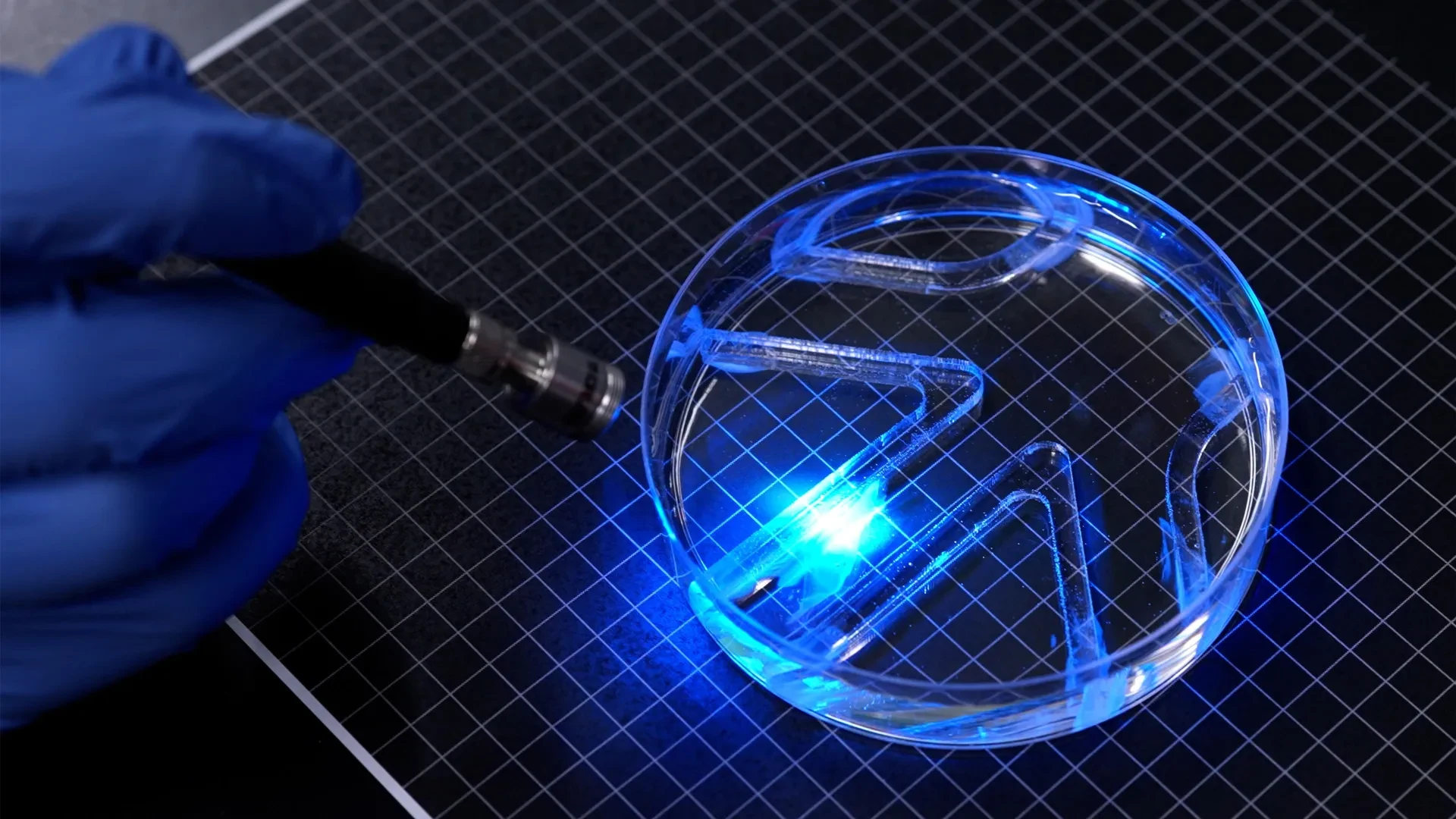
To validate the performance of the completed device, the MIT team placed the swimming robot inside a large petri dish filled with water, featuring a custom-built underwater obstacle course.
Using an external light source manipulated by hand, the researchers selectively illuminated the left or right fin. The targeted light exposure triggered immediate, localized muscle contractions, causing the corresponding fin to flap. By alternating or synchronizing the light pulses, the team successfully steered the robot forward, executed sharp turns, and navigated it flawlessly through the underwater maze.
Supporting Context & Metrics
The intersection of biology and robotics—known as biohybrid robotics—offers unique engineering advantages, but it also presents formidable physical hurdles. Chief among these is fluid dynamics.
The Physics of Aquatic Propulsion
Moving through water requires significantly more mechanical force than moving through air due to fluid density and hydrodynamic drag. For a microscopic robot, overcoming water resistance with soft, biological actuators is an immense engineering feat.
During experimental trials, MIT’s thin biohybrid swimmer covered approximately four times its body length per minute at its maximum sustained speed. While this metric pales in comparison to an elite human Olympic swimmer—who can travel up to 65 body lengths per minute—it places the biohybrid robot on par with the relaxed, energy-efficient cruising speed of a living cow shark navigating the ocean currents. Given that the entire propulsion system is generated by a single, ultra-thin cellular layer, this speed-to-size ratio highlights the remarkable force-generating capacity of engineered skeletal muscle.
Efficiency Comparison: 2D vs. 3D Biohybrid Robots
Prior to this breakthrough, the vast majority of biohybrid swimming and walking robots were constructed as bulky, three-dimensional structures. These traditional devices typically relied on thick chunks of laboratory-grown skeletal muscle, requiring millions of individual cells to fabricate.
While functional, these 3D designs suffer from several critical drawbacks:
- High Manufacturing Costs: Culturing and maintaining millions of cells demands expensive nutrient media, specialized incubator space, and extensive labor.
- Massive Diffusion Limits: In thick 3D tissue constructs, oxygen and nutrients struggle to diffuse to the innermost cells, often leading to necrotic cores where cells die off before the robot can even be deployed.
- Reduced Mechanical Efficiency: Heavy, bulky muscle masses introduce excess inertia and structural drag, reducing the overall propulsion efficiency of the device.
By contrast, the MIT team’s two-dimensional approach utilizes a single, ultra-thin cellular layer. This design virtually eliminates nutrient diffusion barriers, slashes manufacturing costs by dramatically reducing the required cell count, and maximizes energy transfer efficiency directly into the surrounding water.
Official Statements
The implications of this research have drawn widespread attention within the academic and engineering communities. Lead author Ritu Raman emphasized both the mechanical potency of the device and its long-term vision for real-world deployment.
"It takes a lot of force to move through water versus air," noted Ritu Raman, associate professor of mechanical engineering at MIT. "The robot’s quite strong, given its size."
Addressing the broader landscape of biohybrid engineering and why transitioning from bulky three-dimensional structures to streamlined two-dimensional designs is so transformative, Raman elaborated:
"Currently, biohybrid robots from our group and others’ are built from bulky, 3D chunks of lab-grown skeletal muscle that require millions of cells to fabricate," Raman explained. "We believe that biohybrid robots powered by living muscle could one day perform delicate jobs like exploring environments too fragile or unpredictable for conventional hardware, because living tissue is soft, responsive to its surroundings, and can heal itself."
Detailing the mechanics of the dual-fin propulsion system and how the team achieved precise directional control, Raman added:
"You can think of the robot as having two independent muscles. If we shine a light on just one, only that muscle moves. If shining on both, they both flap."
Reflecting on the foundational challenges overcome during the project—specifically moving from microscopic displacements to macroscopic propulsion—Raman recalled the limitations of their earlier work:
"People hadn’t seen this muscle architecture engineered from scratch before. And the cells were moving in multiple directions. But they only moved about 100 microns. From a robotics perspective, their movements were tiny."
The research team behind this publication also included first author Maheera Bawa, alongside contributing researchers Arielle Berman, Laura Schwendeman, Ferdows Afghah, and Seanbiron Johnson. Financial and institutional support for the project was provided in part by the Office of Naval Research, underscoring the potential military and strategic applications of stealthy, soft-bodied aquatic systems.
Future Outlook
While the successful navigation of an underwater maze marks a major milestone, the MIT research team views this study as merely the opening chapter for two-dimensional muscle-powered robotics.
Short-Term Engineering Objectives
The immediate priority for Raman’s laboratory is optimizing the bodily morphology and hydrodynamic profile of the swimmer. Although the current prototype successfully demonstrated locomotion and steering, its overall body shape remains intentionally basic. By refining the aerodynamic and hydrodynamic contours of the gel framework, the engineers aim to significantly increase top-end swimming speeds and maneuverability.
Long-Term Applications: Environmental Monitoring and Beyond
Looking further into the future, the integration of living tissue into robotic systems opens up transformative possibilities that rigid metallic or plastic robots cannot match:
- Fragile Aquatic Ecosystems: Conventional autonomous underwater vehicles (AUVs) driven by metallic propellers and loud electric thrusters can disrupt delicate coral reefs, stir up sediment, and harm aquatic life. Soft, muscle-powered robots move fluidly and silently, blending seamlessly into natural aquatic habitats.
- Self-Healing Infrastructure: Because the primary actuator is living biological tissue, future iterations of these robots could possess intrinsic self-healing properties, repairing micro-tears or fatigue damage autonomously over time.
- Environmental Sensing: Equipped with biological or chemical sensors, swarms of these ultra-thin biohybrid swimmers could be deployed to monitor water quality, detect pollutants in tight subterranean pipe networks, or map delicate underwater caves inaccessible to traditional hardware.
As biohybrid engineering continues to mature, the boundary between machine and organism is steadily dissolving. MIT’s breakthrough proves that nature’s time-tested actuators—muscle cells—can be successfully harnessed, engineered, and directed to perform complex mechanical tasks, setting the stage for a new era of living machines.