

Executive Overview
In the relentless pursuit of blending organic biology with synthetic mechanical engineering, researchers at the Massachusetts Institute of Technology (MIT) have achieved a striking breakthrough. Traditional robotics has long relied on bulky, power-hungry electric motors, heavy metallic frames, and complex mechanical linkages to achieve locomotion. However, a team of MIT engineers has successfully demonstrated that an extraordinarily thin, two-dimensional layer of living muscle tissue can generate enough mechanical force to autonomously propel a swimming robot through water.
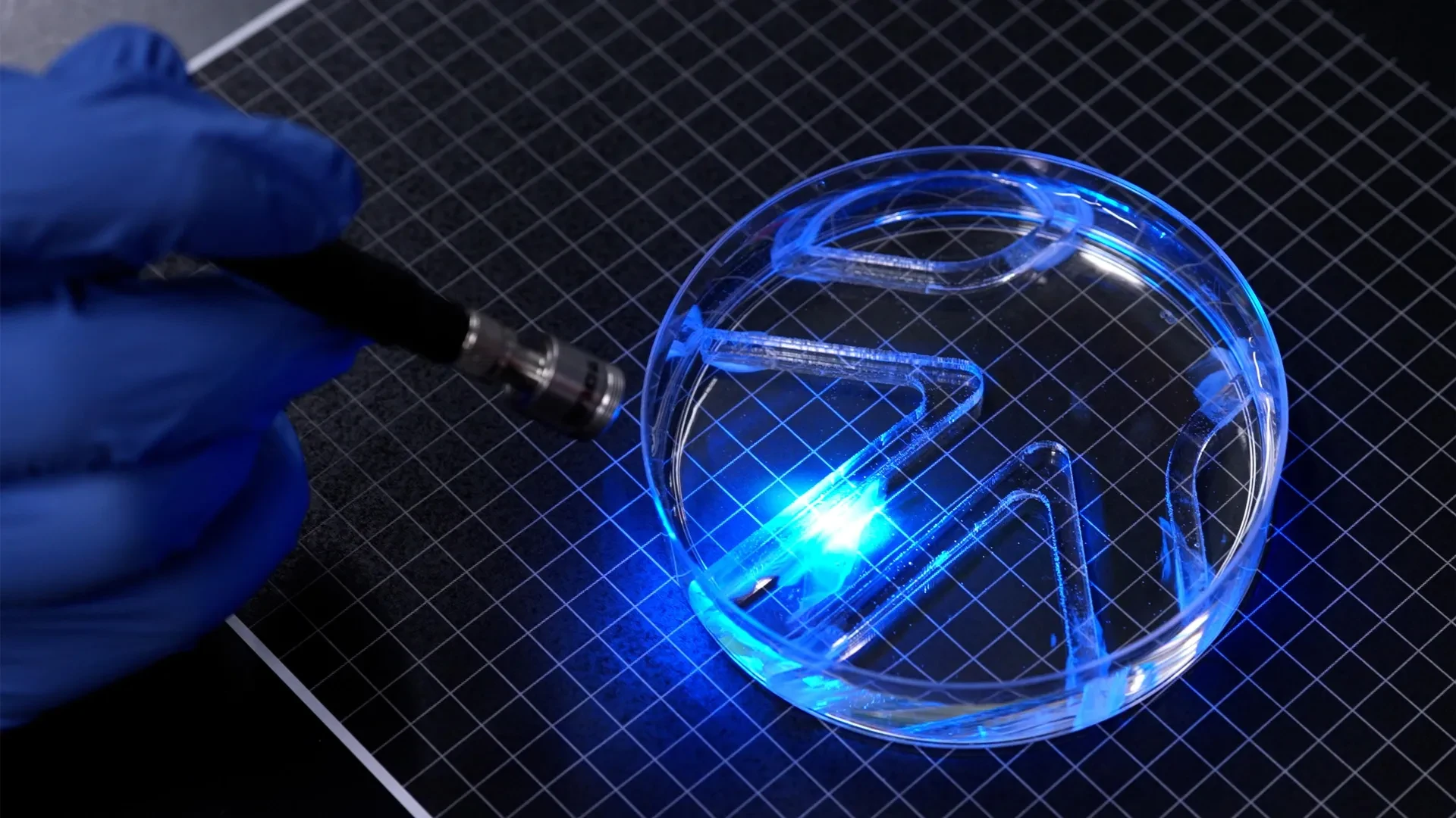
Detailed in a landmark study published in the journal Advanced Functional Materials, this microscopic marvel features a flexible, gum-stick-sized gel framework partitioned into two distinct fins. Each fin is coated with a layer of lab-grown skeletal muscle cells significantly thinner than a human hair. Genetically modified to respond to external light stimuli, these cells contract on command when illuminated, driving the fins to flap and push the device through an aqueous environment.
By selectively pulsing light onto either the left or right fin, the research team can dictate both the speed and directional heading of the robot. In experimental trials, the biohybrid device successfully navigated a complex underwater maze, showcasing precise steering capabilities. At maximum velocity, the robot covered roughly four times its body length per minute. While this pace is modest compared to elite human swimmers—who can reach up to 65 body lengths per minute—it mirrors the unhurried, graceful cruising speed of a cow shark navigating the ocean depths.
Beyond its immediate aquatic performance, this research represents a paradigm shift in biohybrid engineering. Most contemporary biohybrid machines utilize bulky, three-dimensional blocks of lab-grown tissue requiring millions of cells to construct. By scaling down to a micro-thin, two-dimensional architecture, the MIT team has opened the door to drastically lower manufacturing costs, enhanced energy efficiency, and a new generation of soft, responsive, self-healing robots capable of exploring fragile ecosystems where rigid hardware would cause catastrophic damage.
Detailed Chronology
From Artificial Irises to Propulsion: The Evolutionary Timeline
The genesis of this swimming robot traces back to an earlier achievement within the laboratory of Ritu Raman, associate professor of mechanical engineering at MIT. Last year, Raman’s team introduced an artificial muscle disk directly inspired by the intricate mechanics of the human eye’s iris. This foundational project sought to prove that a microscopic layer of living cells could be coaxed into producing precisely controlled, multidirectional movements.
To construct the artificial iris, the engineers patterned a soft gel disk with microscopic grooves extending radially outward from its center and forming concentric circles. When living muscle cells were seeded onto this surface, they naturally migrated into the channels, growing into an organized, ultra-thin layer. Upon illumination, the muscle tissue contracted in multiple directions, causing the disk to stretch and compress dynamically—mimicking the way an iris pupil expands and contracts.
While this iris experiment successfully established that researchers could engineer intricate muscle architectures from scratch, the actual physical displacement was minuscule. The cells only moved about 100 microns. From a practical robotics perspective, the displacement was too small to perform useful work.
Determined to bridge the gap between microscopic cellular twitching and macroscopic robotic locomotion, Raman and her team—including lead author Maheera Bawa, Arielle Berman, Laura Schwendeman, Ferdows Afghah, and Seanbiron Johnson—embarked on a rigorous quest to dramatically scale up the mechanical force generated by the tissue.
Overcoming Structural Limitations
The primary roadblock in the earlier iris project was the choice of substrate: fibrin. While fibrin is an exceptionally soft and biocompatible gel, it proved structurally unstable under the heavy physical loads exerted by contracting muscle cells. The material tended to shrink rapidly, severely limiting the range and force of the resulting movement.
To overcome this hurdle, the MIT engineers systematically investigated three fundamental variables of the gel foundation:
- Chemical composition
- Material stiffness
- The geometric dimensions and profile of the surface grooves
The team hypothesized that by tuning these parameters, they could create an optimized matrix capable of supporting robust muscle growth without succumbing to structural deformation.
Material Optimization and Grooved Architecture
The researchers tested a variety of polymer formulations, stiffness levels, and micro-groove geometries. They compared narrow channels featuring square profiles against longer channels with curved bottoms. Through rigorous microscopic analysis, they discovered that muscle cells aligned significantly better within square-shaped grooves.
This alignment was a critical breakthrough. When muscle cells grow uniformly in the same direction, they naturally fuse into elongated muscle fibers. These cohesive fibers produce far stronger, synchronized contractions than disorganized cell clusters, providing the high-tensile force required to push against water resistance.
Next, the team replaced the problematic fibrin with gelatin methacrylate (GelMA), a versatile hydrogel widely utilized in tissue engineering. By adjusting the chemical formulation of the GelMA, the engineers synthesized supporting films with varying degrees of rigidity. Contrary to the intuition that soft tissues require soft beds, the team discovered that stiffer gels yielded superior results. On firmer GelMA surfaces, muscle cells achieved optimal alignment and generated explosive, high-force contractions.
Thickness and Conditioning
Finding the ideal thickness for the gel film required a delicate balancing act. The engineers determined that a film measuring approximately half a millimeter offered the optimal sweet spot. It was sturdy enough to act as a reliable anchor for the muscle cell layer, yet sufficiently lightweight and flexible to prevent the powerful, contracting tissue from shearing off the surface.
To further enhance performance, the team subjected the engineered tissue to a form of biological "exercise." Before integrating the muscle-coated gel into the final swimming robot, the researchers exposed the cells to repeated pulses of light. This routine stimulation conditioned and strengthened the muscle fibers, priming them for optimal force output once submerged.
Assembly and Maze Navigation
With the gel framework optimized, the muscle tissue strengthened, and the cellular pathways aligned, the researchers assembled the final swimming device. The robot was cast as a thin, gum-stick-sized gel sheet split down the middle to form two independent fins. Both fins were patterned with square-bottomed micro-grooves and seeded with living muscle cells that matured into coordinated, light-sensitive fibers.
The device was then placed inside a large petri dish filled with water, which housed a miniature obstacle course simulating an underwater maze. Using a handheld light source, the researchers independently illuminated the left and right fins. As light hit the targeted cells, the corresponding muscle fibers contracted, causing the fin to flap rhythmically. By dynamically shifting the light beam, the operators steered the biohybrid robot smoothly around obstacles, proving that a micro-thin layer of living tissue could successfully power and navigate an untethered aquatic vehicle.
Supporting Context & Metrics
The Physics of Aquatic Locomotion
Moving through water presents vastly different physical challenges compared to moving through air. Water is roughly 800 times denser and about 50 times more viscous than air, meaning any object attempting to travel through an aquatic medium must overcome substantial hydrodynamic drag.
[Fluid Dynamics Comparison]
+-------------------------+---------------------------------+---------------------------------+
| Metric                  | Water (Aquatic Environment)     | Air (Terrestrial Environment)   |
+-------------------------+---------------------------------+---------------------------------+
| Density                 | ~1,000 kg/m³                    | ~1.2 kg/m³                      |
| Viscosity Resistance    | High                            | Low                             |
| Propulsion Challenge    | Requires high force/drag offset | Requires aerodynamic efficiency |
+-------------------------+---------------------------------+---------------------------------+
As Ritu Raman noted during the study, generating forward momentum in water requires immense mechanical force relative to the object’s physical dimensions. The fact that an ultra-thin, gum-stick-sized biohybrid robot can propel itself through water demonstrates an impressive strength-to-weight ratio for living mammalian tissue.
Quantitative Performance Metrics
To contextualize the robot’s capabilities, researchers evaluated its speed against biological organisms and engineering benchmarks:
- Robot Top Speed: Approximately 4 body lengths per minute.
- Human Swimmer Benchmark: An elite human athlete can cover up to 65 body lengths per minute.
- Comparative Biological Benchmark: Matches the leisurely cruising speed of a cow shark (Hexanchus griseus) gliding through oceanic currents.
- Thickness Profile: Muscle cell layer is substantially thinner than a human hair, supported by a 0.5-millimeter gel film framework.
- Displacement Scale: Overcomes previous micro-scale limitations, jumping from microscopic displacements of 100 microns to macroscopic, maze-navigating propulsion.
Scaling and Cost Efficiency in Biohybrid Systems
Traditional biohybrid robots are hampered by their reliance on massive, three-dimensional tissue scaffolds. Fabricating these structures demands millions of laboratory-grown cells, which drives up production costs and introduces logistical bottlenecks in incubation and maintenance.
By transitioning to a two-dimensional, micro-thin architecture, MIT’s approach slashes the required cell count. This reduction in biological material not only lowers manufacturing overhead but also enhances internal nutrient diffusion, preventing the necrotic cores that often plague thick, 3D bio-constructs. Consequently, these streamlined designs offer a scalable blueprint for mass-producing biohybrid machinery.
Official Statements
The research team emphasized both the fundamental mechanics and the broader philosophical implications of marrying living biology with robotics.
Dr. Ritu Raman, associate professor of mechanical engineering at MIT and the principal investigator of the study, highlighted the unique advantages of utilizing organic tissue over conventional synthetic actuators:
"It takes a lot of force to move through water versus air. The robot’s quite strong, given its size."
"Currently, biohybrid robots from our group and others are built from bulky, 3D chunks of lab-grown skeletal muscle that require millions of cells to fabricate. We believe that biohybrid robots powered by living muscle could one day perform delicate jobs like exploring environments too fragile or unpredictable for conventional hardware, because living tissue is soft, responsive to its surroundings, and can heal itself."
Addressing the iterative design process and the transition from microscopic observation to macroscopic application, Raman reflected on the limitations of their previous work:
"People hadn’t seen this muscle architecture engineered from scratch before. And the cells were moving in multiple directions. But they only moved about 100 microns. From a robotics perspective, their movements were tiny."
Detailing the mechanics of the dual-fin navigation system, Raman explained:
"You can think of the robot as having two independent muscles. If we shine a light on just one, only that muscle moves. If shining on both, they both flap."
Looking toward practical deployment, Raman emphasized the tuning parameters inherent in tissue engineering:
"For engineering any type of tissue, it’s known that these are knobs you can tune. And we wanted to optimize all these parameters to support live muscle cells."
Future Outlook
Roadmap for Next-Generation Biohybrid Systems
While the current iteration of the MIT swimming robot successfully demonstrates the viability of micro-thin muscle propulsion, its structural morphology remains intentionally simple. The primary objective of this initial phase was to establish proof-of-concept: proving unequivocally that an exceptionally thin sheet of engineered muscle could generate sufficient force to swim and steer.
With that milestone achieved, the research team has outlined an ambitious roadmap for future development.
- Velocity Optimization: The immediate engineering priority is refining the body design, hydrodynamic profile, and fin kinematics to dramatically increase swimming speeds. By tweaking the aspect ratio of the fins and optimizing light-pulse frequencies, the team aims to push the robot well beyond its current pace of four body lengths per minute.
- Autonomous Sensory Integration: Currently, the robot relies on an external, manual light source to guide its movements through a maze. Future iterations could incorporate onboard microprocessors, microscopic organic sensors, or chemical-gradient detectors, allowing the robots to autonomously respond to environmental cues without human intervention.
- Complex Multi-Actuator Systems: Expanding beyond simple two-fin propulsion, engineers plan to design multi-segment biohybrid bodies capable of complex, undulating swimming gaits reminiscent of fish or aquatic mammals, further enhancing maneuverability in turbulent waters.
Real-World Applications and Environmental Monitoring
The long-term vision for muscle-powered robotics extends far into practical, high-value field applications. Conventional submersibles—driven by metallic propellers, spinning gears, and rigid hulls—pose inherent risks to delicate aquatic habitats. They can crush fragile coral reefs, disrupt sensitive marine life through acoustic noise pollution, and leave toxic chemical footprints if batteries or lubricants leak.
In contrast, biohybrid robots constructed from soft gels and living tissue offer a harmonious alternative:
- Aquatic Ecosystem Monitoring: Deployed in fragile coral reefs, kelp forests, or pristine freshwater lakes, these soft swimmers can blend into the background, operating silently without disturbing local fauna.
- Hazardous Infrastructure Inspection: Capable of navigating tight, convoluted spaces—such as submerged industrial pipelines, narrow drainage systems, or flooded subterranean caverns—muscle-powered bots can inspect structural damage where rigid hardware would wedge or break.
- Self-Healing Resilience: Because the primary driving mechanism is living tissue, these devices possess intrinsic biological maintenance properties, opening the theoretical possibility of minor self-repair in isolated operational zones.
Supported in part by funding from the Office of Naval Research, this research marks a critical milestone in robotics. By harnessing the quiet, efficient power of living muscle cells, MIT engineers have moved one step closer to a world where machines are no longer built merely to interact with nature, but are seamlessly integrated with it.